Article

Modified U-Tube for Ruling out Naked DNA Transfer during Conjugation and Application in Antibiotic Resistance Genes Transfer Research

1
School of Environment, Tsinghua University, Beijing 100084, China
2
School of Energy and Environmental Engineering, University of Science and Technology Beijing, Beijing 100083, China
3
School of Water Resources and Environment, China University of Geosciences (Beijing), Beijing 100083, China
*
Authors to whom correspondence should be addressed.
Water 2018, 10(10), 1313; https://doi.org/10.3390/w10101313
Submission received: 6 July 2018 / Revised: 18 August 2018 / Accepted: 18 September 2018 / Published: 22 September 2018
(This article belongs to the Section Water Quality and Contamination)

Abstract

:
Antibiotic resistance is currently a major global public health issue. In particular, the emergence and transfer of antibiotic resistance genes (ARGs) is a matter of primary concern. This study presented a method for ruling out the transfer of naked DNA (plasmid RP4 lysed from donor cells) during the cell-to-cell conjugation, using a modified “U-tube”. A series of gene transfer assays was conducted in both flask and modified U-tube, using Pseudomonas putida KT2440 (P. putida (RP4)) harboring the RP4 plasmid as the donor strain, Escherichia coli (E. coli, ATCC 25922) in pure culture as sole recipient, and bacteria from reclaimed water microcosms as multi-recipients. The verification experiments showed that the U-tube device could prevent direct contact of bacteria without affecting the exchange of free plasmid. In the experiments involving a sole recipient, the transconjugants were obtained in flask samples, but not in modified U-tube. Furthermore, in experiments involving multi-recipients, transfer of naked DNA in the modified U-tube accounted for 5.18% in the transfer frequency of the flask transfer experiment. The modified U-tube proved to be useful for monitoring the interference of naked DNA in the research of conjugative transfer and calculating the exact conjugative transfer rate. This device is identified as a promising candidate for distinguishing different gene transfers in practical application because of its convenient use and easy and simple manufacture.

Graphical Abstract

1. Introduction

In recent years, antibiotic resistance has received increasing attention as a major global public health issue [1,2,3]. One of the greatest concerns about the presence of antibiotics in the environment is the emergence and dissemination of antibiotic resistant genes (ARGs). To illustrate the severe risk of ARGs, the World Health Organization named the World Health Day in 2011 as “Antimicrobial Resistance and Its Global Spread” day. Progressively increasing ARGs including genes that confer resistance to sulfonamides, β-lactams, trimethoprim, tetracyclines, aminoglycosides, macrolides, and quinolones [4,5,6,7,8,9] have been detected in various environments. ARGs can transfer among bacteria by vertical gene transfer (VGT) and horizontal gene transfer (HGT) [10,11]. HGT facilitates the direct transfer of DNA between microorganisms, and it is regarded as the major evolutionary force that shapes microbial diversity [12]. There are three major mechanisms for HGT [13]: conjugation (genes transfer through cell-to-cell contact), natural transformation (uptake of exogenous DNA from the environment), and transduction (delivery of genetic material through phage).
Although conjugative transfer is considered to be the main mechanism that contributes significantly to the dissemination of ARGs [14], the gene transfer may also occur through transformation or transduction [15]. However, studies about HGT in bacteria [16], soil [17], wastewater [18], human intestines [19], and even plants [20] were more focused on the conjugative transfer and did not clearly distinguish natural transformations from conjugative transfer. Streptococcal species have been shown to exchange conjugative transposons via transformation in addition to conjugation [21]. Similarly, a great abundance of ARGs outside of cells (extracellular ARGs, eARGs) has been proven to be present in the natural environment [22,23], which originate from the lysis of dead antibiotic resistance bacteria (ARB) or are secreted by living ARB. Thus, due to the presence of eARGs, dissemination of ARGs is easily possible via natural transformation in certain environments [24,25]. Therefore, eARGs might cause a serious risk to ecosystem or even to human health, which cannot be ignored.
Recently, more and more studies on HGT have been extensively devoted to conjugation. However, the most commonly studied conjugation [26,27] might always involve the transfer of eARGs (i.e., transformation). Therefore, investigation of the transfer of eARGs can aid in better understanding and clarification of their contribution to the dissemination of ARGs. In previous studies, naked DNA (extracted from donor bacteria) was directly added to the recipient system and used as control for ruling out the natural transformation in the conjugative transfer process [28]. However, this method does not accurately estimate the interference of transformation. This is attributed to the fact that the donor bacteria cannot all die or dissolve and release all the plasmids; thus, determination of a precise gene transfer rate is crucial for ARGs risk assessment. Therefore, it is important to discriminate transfer of naked DNA from conjugation.
In this study, a modified apparatus, namely U-tube, was introduced, which was used to eliminate the interference of naked plasmid transfer (lysed from donor cells) with conjugative transfer. This system could efficiently distinguish transfer caused by naked DNA, i.e., transformation from conjugation in HGT without plasmid extraction in donor cells and calculate the transfer frequency accurately. Most of all, the device is convenient to operate, easy to manufacture, and reusable.
Bacteria tagged with fluorescent protein(s) were used herein to detect the gene transfer [29]. The numbers of donors, recipients, and transconjugants (or transformants) were evaluated by flow cytometry (FCM) and confocal laser scanning microscopy (CLSM).

2. Materials and Methods

2.1. Bacterial Strains

The donor strain was Pseudomonas putida KT2440 (P. putida (RP4)) [30], chromosomally tagged with mCherry red fluorescence and lacIq, and harboring the RP4 plasmid [31], which carries the gfp gene (RP4::Plac::gfp) and confers resistance to kanamycin. The expression of gfp was repressed in the donor cell; however, rfp was constitutively expressed. The donor strain was grown in lysogeny broth (LB) medium containing kanamycin (50 mg·L−1), and shaken overnight at 30 °C.
Figure 1 illustrates the principle of gene transfer of strain [29]. The donor strain showed red fluorescence with mCherry tagged on chromosome. If the plasmid transfers to a non-fluorescent recipient strain, that strain acquires green fluorescence, while the donor strain shows red fluorescent tags. Thus, donors, recipients and transconjugants (or transformants) can be distinguished.
Escherichia coli (E. coli, ATCC 25922) bacteria in pure culture were used as sole recipient in this study. It is sensitive to antibiotics; and it did not show fluorescent protein expression. The recipient was grown in LB medium at 30 °C by shaking overnight.
Bacterial system was prepared from reclaimed water microcosm by following the method described by Luo et al. [32], and it was used as multi-recipient. The reclaimed water was collected at the reclaimed water recharge point of Chaobai River, Beijing in September 2017. The reclaimed water was produced from the wastewater treatment plant and further treated in a membrane bio-reactor. Samples were collected in sterile brown glass bottles, stored at 4 °C, and then further processed immediately upon arrival in the laboratory. Samples were stored in refrigerator at 4°C for 3 h. The supernatant was collected and supplemented with LB media (v:v = 1:1), and then incubated overnight at 30 °C on a shaker.
In this study, “transconjugant” is defined as a cell that has received the fluorescence-conferring plasmid in cell-to-cell conjugation; “transformant” is a cell that acquired RP4 plasmid in RP4 transfer assay; and a “recipient” is a cell that has not taken up the fluorescence-conferring plasmid.

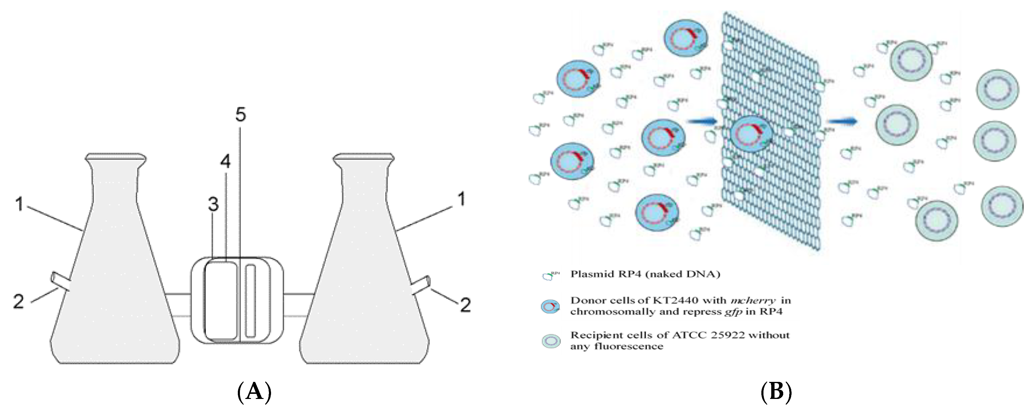
2.2. Modified U-Tube Device

The U-tube system was modified from the device mentioned in literature report [33], which showed that conjugation needs cell-to-cell contact. The U-tube mentioned in the original reference was a glass tube. Filter was sintered into the tube, forming a monolithic U-tube. The pore diameter was difficult to control, and the pores could easily be blocked. In this study, modified U-tube containing two 250 mL culture flasks each with a suction filter joint on the side was used. The two flasks were connected through a flange containing two silicone gaskets and a 0.22 μm filter membrane. Schematic illustration of the apparatus is shown in Figure 2A.
The filter membrane used in this study was a cellulose nitrate membrane, which could prevent exchange of cells between the flasks without interfering with the exchange of other molecular substances in the culture medium, including plasmids. The principle of operation is shown in Figure 2B. The diameter of bacteria is about 1 μm, so they are prevented from passing from one chamber to the other through the filter, whereas macromolecules and micromolecules could be transferred through the membrane pore. The pore size of the membrane was selected according to the size of the microorganisms studied. The suction filter joint was used to connect the flasks to a vacuum pump, and the gaskets acted as a seal to avoid leakage. The leakage tests were performed using water and sterile LB medium vs P. putida, respectively. Details of these tests were listed in the Supplementary Materials (Text S1 and Figure S1).
The two culture flasks were inoculated with the donor and recipient strains, which were named as donor and recipient flask, respectively. During the experiment, the exchange of liquid between the two flasks of the U-tube was assisted by the vacuum pump. The flow rate between the two flasks was 3 mL·min−1. Furthermore, the U-tube system was placed on a shaker (160 rpm) until the completion of the experiment.

2.3. Verification of U-Tube Performance

The donor harboring the plasmid RP4 was grown overnight at 30 °C in LB with 50 mg·L−1 kanamycin. RP4 was extracted in the cultured P. putida (RP4) using an E.Z.N.A. Plasmid Mini kit I (Omega Bio-tek, Norcross, GA, USA) according to the manufacturer’s instructions (the final concentration was 5 ng·μL−1). To verify the performance of the U-tube, the naked plasmid RP4 was inoculated in the donor flask of the U-tube, while sterile LB medium was injected in the recipient flask and named as Test 1#. Test 2# was set as follows: P. putida (RP4) (washed three times with phosphate-buffered saline (PBS) and OD600 was adjusted to around 0.5) was taken in the donor flask and E. coli in the recipient flask of the U-tube. The system was pumped to assist the exchange of liquid between the two flasks of the U-tube. Experiments in flasks with RP4 vs LB, and P. putida (RP4) vs. E. coli were also set up with the test numbers of 3# and 4#, respectively. The experimental settings were summarized in Table 1.

2.4. Gene Transfer Experiment

The donor strain and the recipient strain (E. coli and microcosm) were cultured, respectively, as described earlier, and grown overnight [34]. To preserve the dying and lysing cells, both the donor and recipient strains used in modified U-tube were adjusted to OD600 nm of ~0.5 with LB without washing with PBS. Donor and recipient strains were then inoculated, respectively, in the donor and recipient flasks of the U-tube with the volume of 80 mL (volume ratio 1:1). Furthermore, the system was pumped to assist the exchange of liquid between sides of the membrane, and the apparatus was left shaking for 3 h at 30 °C.
Cell-to-cell conjugations were also set up in 250 mL flasks containing the donor and recipient strains with volume ratio 1:1 (80 mL, respectively). The donor cells were washed three times with PBS to remove any trace antibiotics and metabolites [35,36], and then suspended in a small volume of LB. The OD600 nm of the culture was adjusted to around 0.5. The recipient cells were also treated with PBS, and then adjusted to OD600 nm of ~0.5. These tests were numbered as 5#–8# as summarized in Table 1.
Donor and recipient strains were further cultured in separate 250 mL flasks as controls, with test numbers of 9#, 10#, and 11#. All the genes transfer tests were conducted over 3 h, which could limit bacterial growth and selection during the experimental phase. Bacterial growth would result in ambiguous data if allowed to occur over a long period [35].

2.5. Sampling and Detection of the Gene Transfer

In the verification tests, samples in the U-tube and flask were sampled at 15, 60, and 180 min, and then the polymerase chain reaction (PCR) was performed for the presence of traF gene (indicator for RP4) and aphA gene (resistant gene to kanamycin on RP4) [37]. Samples of experiments with P. putida (RP4) as donor were assessed on plates containing 50 mg·L−1 kanamycin for the cell transfer. Samples of gene transfer both in U-tube and flask at 180 min were visualized by FCM and CLSM for the presence of transconjugants (or transformants). The details of PCR, FCM and CLSM are described in Text S2 and S3.

3. Results and Discussion

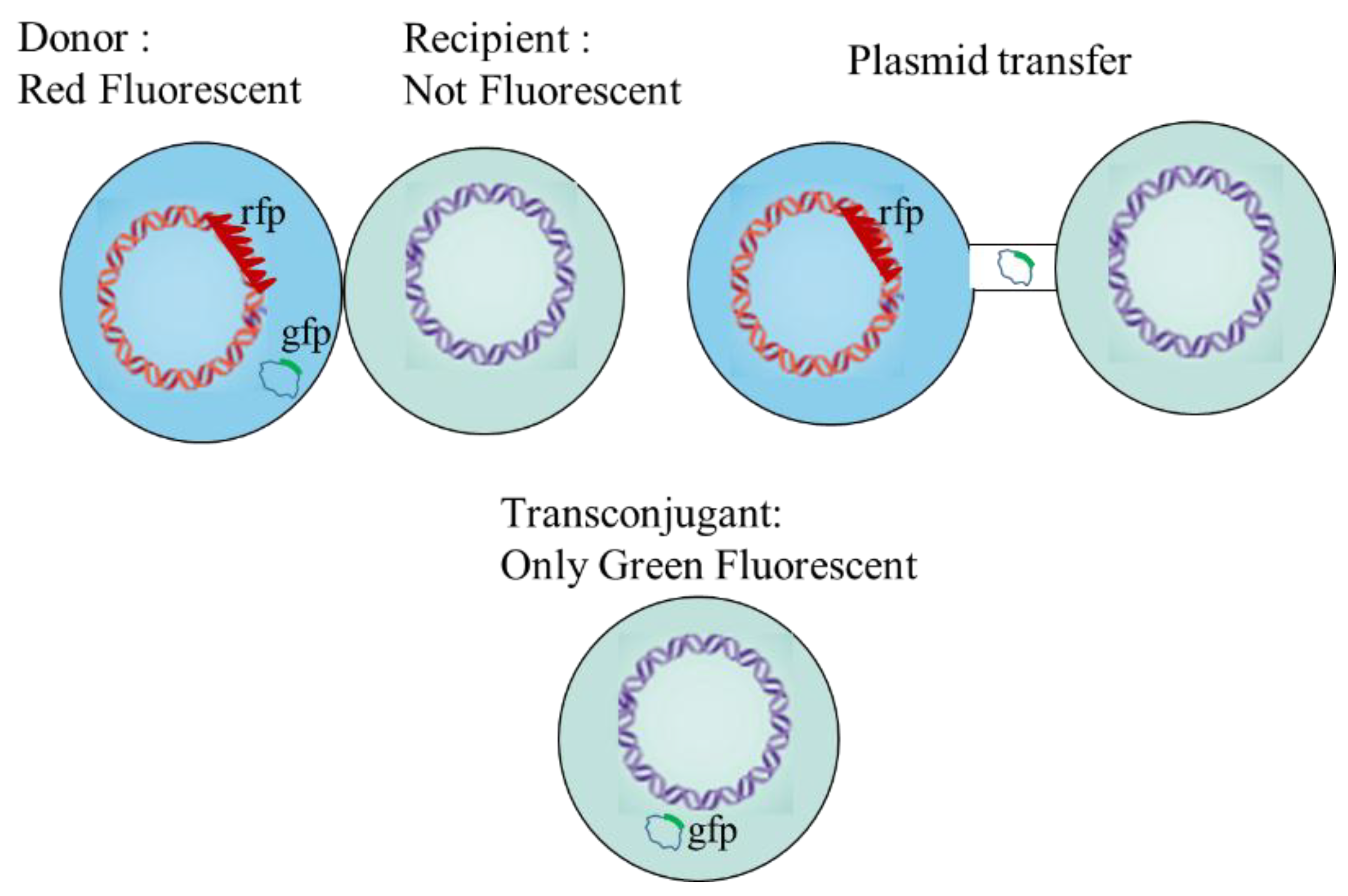
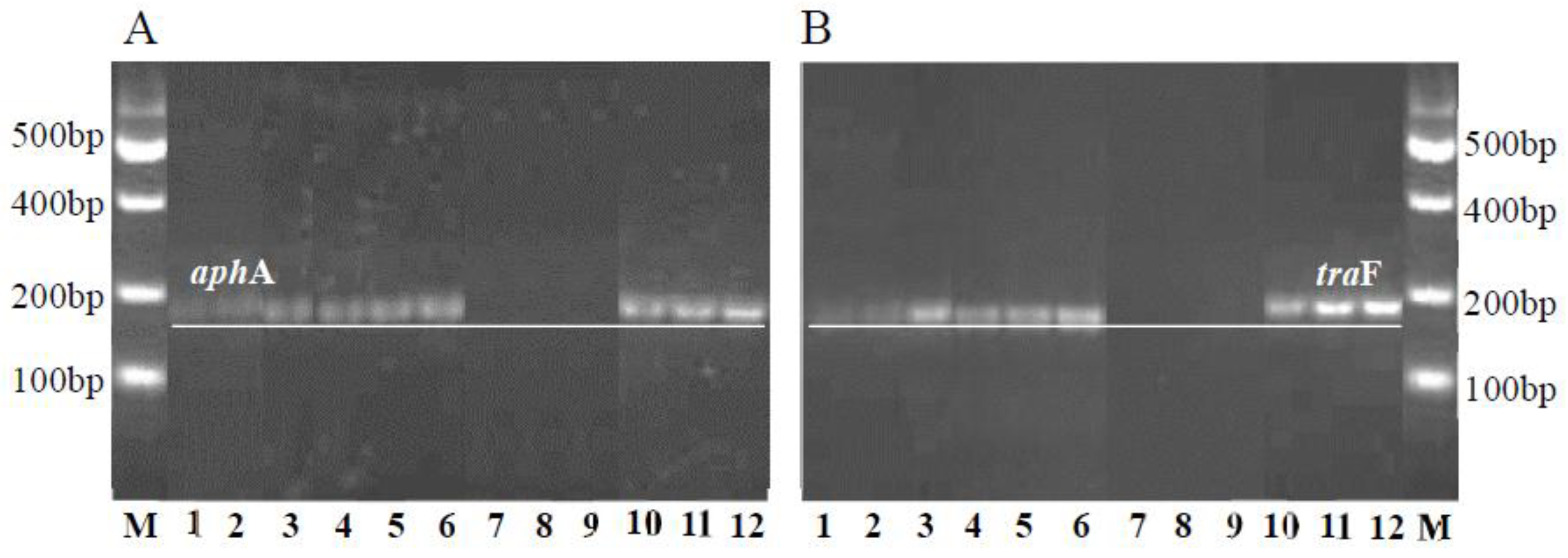
3.1. Transfer of Plasmid and Donor Strain in the U-Tube

All samples for verification tests were subjected to PCR analysis to test the presence of plasmid RP4 (i.e., by detecting genes traF and aphA) (Figure 3). Figure 3 exhibits the presence of bands of genes traF and aphA in recipient samples of Test 1#. The concentration of RP4 increased with the time of experiment. Moreover, the bands on the gel also become clearer with time (the traF gene acid sequences of samples at 180 min are shown in Figure S2). However, no band was detected for recipient flask of U-tube when P. putida (RP4) was inoculated in the donor flask in Test 2#. In other words, plasmid RP4 could penetrate the recipient flask of the U-tube, while P. putida (RP4) could not. This indicates that naked DNA from lysis donor cell could enter the recipient flask, because of its smaller size than that of the membrane (0.22 μm). Selective plates were used to detect the transconjugants in experiments with P. putida (RP4) as donor in U-tube and flask, respectively. By adding kanamycin with a concentration of 50 mg·L−1, only cells that had acquired the resistance gene were detectable and the results are listed in Table 2. The second column shows the total number of colonies forming units (CFU) of P. putida (RP4) (the donor) isolated in the donor flask of the U-tube in Test 2# after 180 min. The number of CFU observed in test 4# increased with time. However, no colony was observed for the samples present in recipient flask of U-tube (Test 2#), indicating the absence of occurrence of conjugation or transformation. No E. coli on the recipient side of the U-tube had taken up RP4 because of the lack of cell-to-cell contact [13]. Therefore, the modified U-tube could prevent the direct contact of cells without affecting the exchange of naked plasmids. It can be used to measure the transfer caused by eARGs, which can pass through 0.22 μm filters. However, column “4#” not only shows transconjugants, but also donor cells. As a confirmation step, FCM and CLSM were used in gene transfer experiments to sort and identify donor, recipient and transconjugant (or transformants) cells by fluorescence.

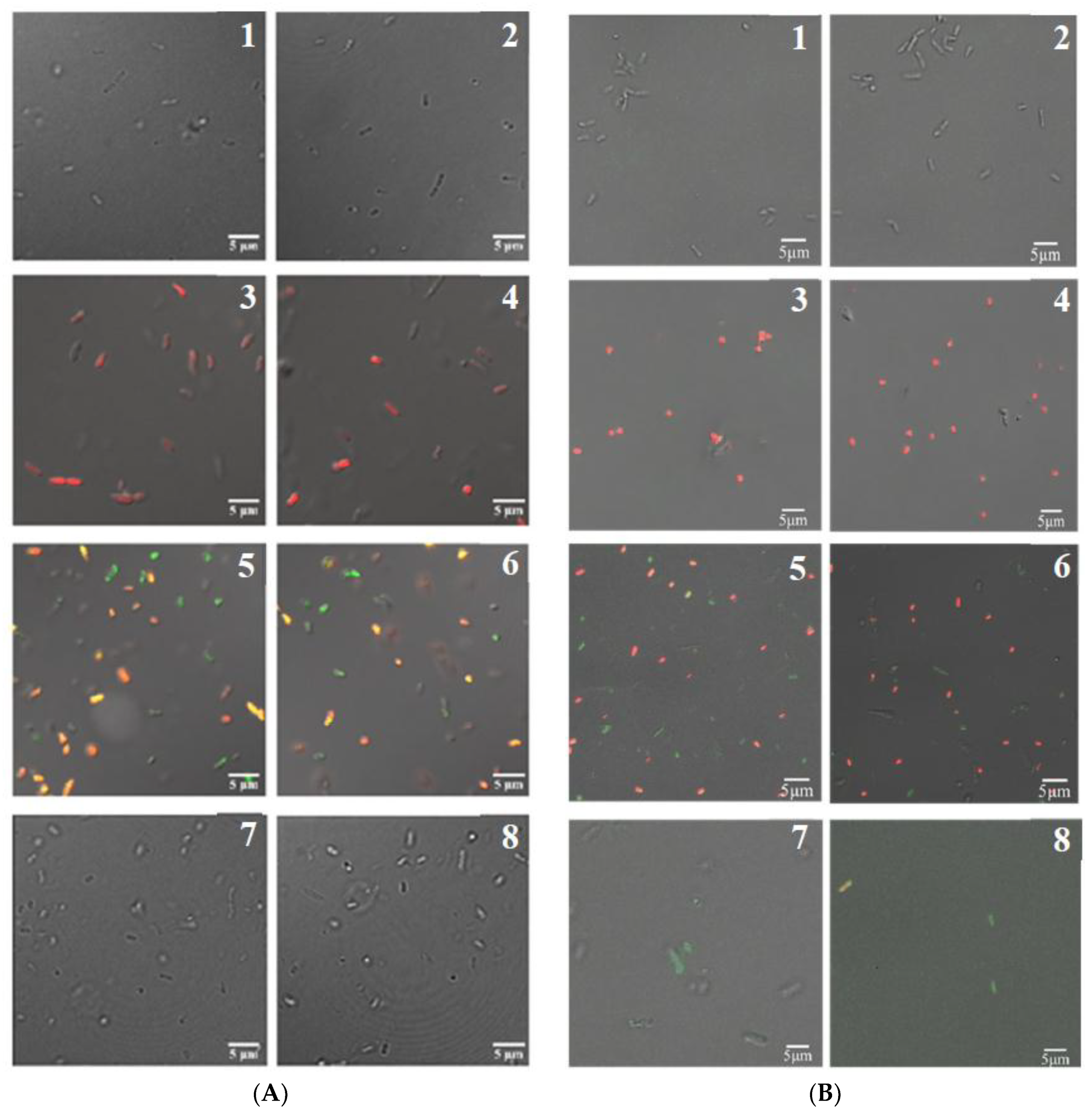
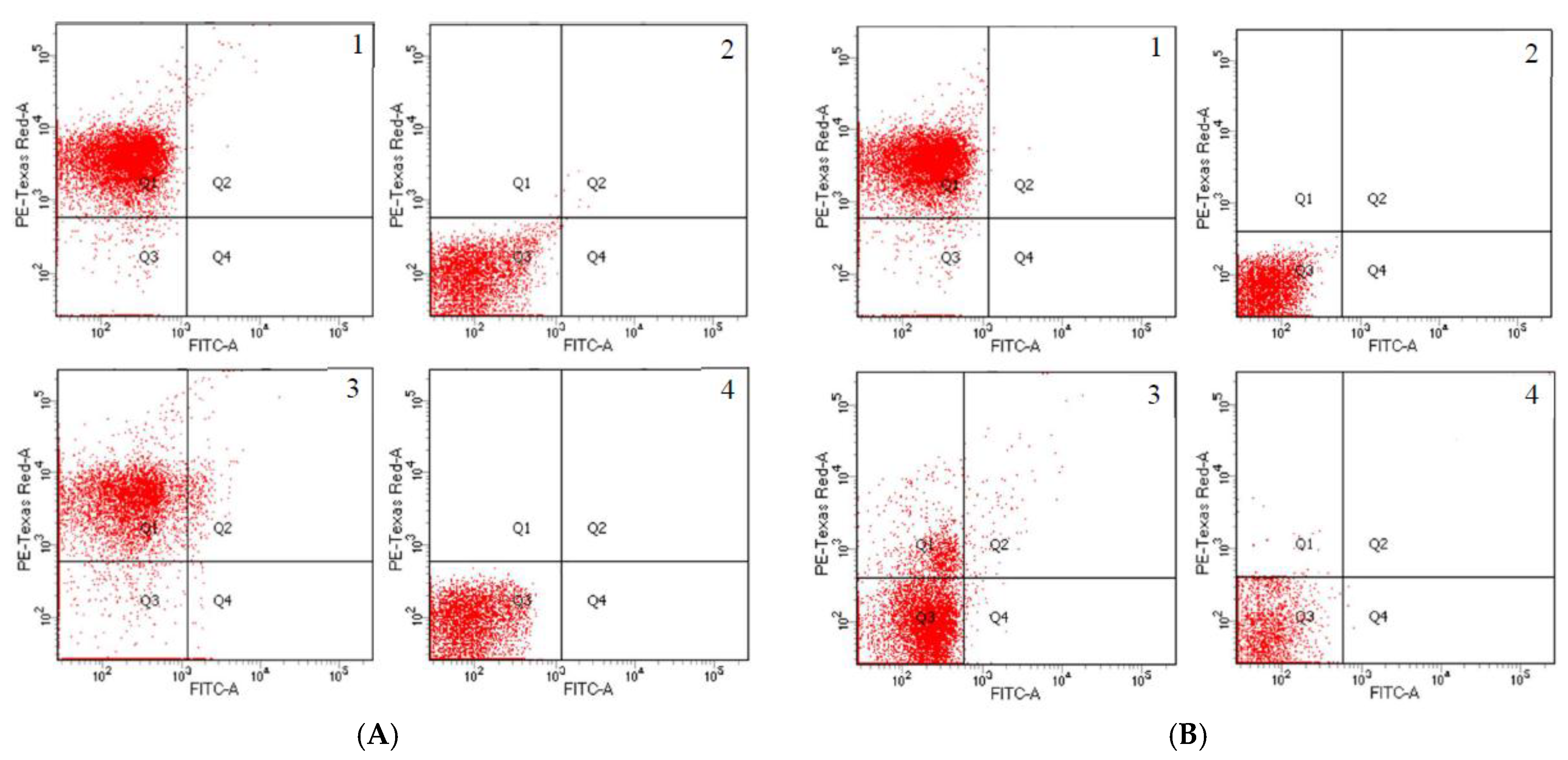
3.2. Detection of Transconjugants in Gene Transfer Experiment

In this study, donor of P. putida (RP4) expresses rfp and represses gfp, while the recipients were not fluorescent. Thus, if the final transconjugants (or transformants) express gfp, it indicates successful gene transfer [38]. The expression of gfp in samples of experiments with E. coli as recipient was observed by CLSM (Figure 4A1–A8). When the experiment progressed for 180 min, only E. coli cells were present; however, no fluorescence was observed for samples of test 5# (Figure 4(A7,A8)). Nonetheless, the green fluorescence increased in flask experiment accompanied by some yellow fluorescence in test 7# (Figure 4A5,A6). The yellow fluorescence obtained in test 7# indicated that the donor cell was right on the stage of losing RP4. At this stage, the RP4 could express green fluorescence without being inhibited. However, the donor cell expresses red fluorescence. When donor cells expressed red and green fluorescence simultaneously, yellow fluorescence was obtained. The same results can also be obtained from Figure 5A3 and 5B3, in which double positive of rfp and gfp events are observed in zone Q2. Fluorescence advantageously provided the transfer information in complex community structures, which were shown in Figure 4B1–B8. Transconjugants in microcosm recipient experiments were detected both in the U-tube samples and flask samples (Tests 6# and 8#). Green fluorescence spread throughout the flask samples (Test 8#), while recipient flask of U-tube had only a few transconjugants (Test 6#). Compared to E.coli, microcosms with more bacteria can accept RP4 secreted from P. putida (RP4), which may be due to the strong species dependence for plasmid [39]. Genera in Proteobacteria and Firmicutes were found to engage in plasmid transfer more easily [36]. Therefore, the complex bacteria communities in reclaimed water resulted in microcosm, which could increase the uptake and incorporation of exogenous DNA into the genome to complete the transformation process.
The dot plots (Figure 5A) also show the same trend for E. coli recipient experiment, with the gfp events being 43 cells at 180 min in flask samples; however, no gfp events were acquired in the U-tube samples. Meanwhile, Figure 5B exhibits that in microcosm experiments the gfp events in flask and U-tube are 42 and 3 cells, respectively. The plasmid transfer frequency obtained from the proportion of gfp to rfp (T/D) [12,40] in Test 7# was 5.30 × 10⁻³, which is higher than that in natural condition (<10−5) [41]. The nutritional level in natural environments is poorer than that in pure culture condition, which might restrict the microbial growth or even the HGT mechanisms [42]. The transfer frequency of transconjugant divided by the recipients (total cells minus rfp) (T/R) [43] in Test 7# was 2.27 × 10⁻³, which is consistent with previous reports [44]. However, in microcosm experiments, the gene transfer rate of Test 8# was 5.87 × 10⁻³. The number of donor cell and the ratio of donor/recipient in the two types of recipient experiments (Tests 7# and 8#) were significantly different (p < 0.01) (Table S2). In fact, the gene transfer rate depends on the density of recipient and donor, environmental factors, opportunity to be in contact, etc. When donor/recipient ratio is significantly small, every donor cell has high opportunity to contact with recipient cells [36]. Therefore, the frequency of transfer in Test 8# was higher than that in Test 7#. Moreover, the gene transfer of T/R in Test 6# was 3.04 × 10⁻³, which accounted for 5.18% of the rate in Test 8#. For the dissemination of ARGs, this ratio could not be ignored.
Owing to the lack of naturally competent cells [45], which were necessary for plasmid transformation [46], transconjugants were not detected in the U-tube experiment of E. coli recipient. Natural transformation was not observed when the donor plasmid RP4 concentration was 5 μg·mL−1, when the concentration was up to 50 μg·mL−1, the transfer frequency was only of the order of 10−9 [32]. The RP4 concentration used in this study was only 5 ng·μL−1. The RP4 concentration transferred into the recipient flask should be lower than 5 ng·μL−1, which was too low for transformation. The microcosm recipient cultured with reclaimed water might be associated with some factors, such as antibiotics, nutritional status, or other organic materials that could induce competence in many species of bacteria [47,48].
In this study, naked plasmid (eARGs) could be exchanged between the two arms of modified U-tube, while P. putida (RP4) (iARGs) could not be. In E. coli recipient experiments, green fluorescence was not obtained in recipient flask of U-tube. When the recipient strains changed to microcosm recipient (bacteria from reclaimed water microcosm), the green fluorescence transconjugants (or transformants) were obtained not only in flask experiment but also in the U-tube, as shown in Figure 4B. The changes in fluorescence are also shown in Figure 5. Green fluorescence is not detected in Figure 5A4. However, slight green fluorescence is observed in Figure 5B4. Moreover, cells in Zone Q4 of Figure 5B3 were transconjugants of Test 8#, which were more than that in Test 7# (Figure 5A3). Zone Q1 of Figure 5B4 exhibits some red fluorescence. This is possibly attributed to the transfer of some chromosome DNA tagged rfp from dead cells into the recipient flask, followed by the completion of the transformation with reclaimed water microcosm. However, whether they were transformation cells from the rfp chromosomal or not, still require further research. In other words, the modified U-tube can be used to eliminate the transformation effects produced by cell lysis in the research of conjugative transfer when conditions are appropriate [46]. Compared to the device used by Davis [33], the U-tube modified in this study can be separated and assembled anytime. Moreover, it can also can be sterilized and reusable. The arms designed as flasks are easy for sampling. The filter can be changed to different pore sizes according to the research material.
However, the conclusion drawn above was based on the assumption that washing with PBS could eliminate eDNA. Owing to the randomness of the sampling and insensitivity of the detection methods, the effectiveness of methods eliminating the free DNA should be studied in further research.

4. Conclusions

The main objective of this study was to develop a method to distinguish conjugation and naked DNA transfer (or even transformation and transduction) in one system. The U-tube was designed using a 0.22 μm membrane to separate the chambers to achieve this purpose. This membrane allows plasmids and other Mobile Genetic Elements (MGEs) to move between the chambers but does not allow movement of intact bacterial cells. Selective media, PCR, CLSM and FCM were used to demonstrate the performance of the U-tube in conjugation and plasmid transfer experiments. In verification experiments, plasmid RP4 was observed in all samples except in the recipient flask of the U-tube when P. putida (RP4) was in the donor flask, which verified that the membrane of the U-tube could prevent cell-to-cell contact. Then the transconjugants (or transformants) were obtained in microcosm experiment in U-tube, which indicates that the modified U-tube can be successfully used to distinguish the plasmid transfer (or transformation effects) from conjugation and provide an exact conjugative transfer date.

Supplementary Materials

The Supplementary Materials are available online at https://www.mdpi.com/2073-4441/10/10/1313/s1.

Author Contributions

Conceptualization, N.Z.; Methodology, N.Z. and B.L.; Validation, X.M., L.H. and F.M.; Formal Analysis, N.Z.; Writing-Original Draft Preparation, N.Z.; Writing-Review & Editing, M.L.; Project Administration, X.L.; Funding Acquisition, X.L.

Funding

This research was funded by the National Natural Science Foundation of China, grant number 51378287.

Acknowledgments

We thank Barth F. Smets, Technical University of Denmark, for the kind gift of Pseudomonas putida KT2440.

Conflicts of Interest

The authors declare no conflict of interest.

References

  1. Levy, S.B.; Marshall, B. Antibacterial resistance worldwide: Causes, challenges and responses. Nat. Med. 2004, 10, S122–S129. [Google Scholar] [CrossRef] [PubMed]
  2. Spellberg, B.; Guidos, R.; Gilbert, D.; Bradley, J.; Boucher, H.W.; Scheld, W.M.; Bartlett, J.G.; Edwards, J., Jr. The epidemic of antibiotic-resistant infections: A call to action for the medical community from the Infectious Diseases Society of America. Clin. Infect. Dis. 2008, 46, 155–164. [Google Scholar] [CrossRef] [PubMed]
  3. Wright, G.D. Antibiotic resistance in the environment: A link to the clinic? Curr. Opin. Microbiol. 2010, 13, 589–594. [Google Scholar] [CrossRef] [PubMed]
  4. Chen, C.; Li, J.; Chen, P.; Ding, R.; Zhang, P.; Li, X. Occurrence of antibiotics and antibiotic resistances in soils from wastewater irrigation areas in Beijing and Tianjin, China. Environ. Pollut. 2014, 193, 94–101. [Google Scholar] [CrossRef] [PubMed]
  5. Graham, D.W.; Knapp, C.W.; Christensen, B.T.; McCluskey, S.; Dolfing, J. Appearance of β-lactam resistance genes in agricultural soils and clinical isolates over the 20th century. Sci. Rep. 2016, 6, 21550. [Google Scholar] [CrossRef] [PubMed] [Green Version]
  6. Huang, H.; Chen, Y.; Zheng, X.; Su, Y.; Wan, R.; Yang, S. Distribution of tetracycline resistance genes in anaerobic treatment of waste sludge: The role of pH in regulating tetracycline resistant bacteria and horizontal gene transfer. Bioresour. Technol. 2016, 218, 1284–1289. [Google Scholar] [CrossRef] [PubMed]
  7. Muziasari, W.I.; Managaki, S.; Pärnänen, K.; Karkman, A.; Lyra, C.; Tamminen, M.; Suzuki, S.; Virta, M. Sulphonamide and trimethoprim resistance genes persist in sediments at Baltic Sea aquaculture farms but are not detected in the surrounding environment. PLoS ONE. 2014, 9, e92702. [Google Scholar] [CrossRef] [PubMed]
  8. Rodriguez-Mozaz, S.; Chamorro, S.; Marti, E.; Huerta, B.; Gros, M.; Sànchez-Melsió, A.; Borrego, C.M.; Barceló, D.; Balcázar, J.L. Occurrence of antibiotics and antibiotic resistance genes in hospital and urban wastewaters and their impact on the receiving river. Water Res. 2015, 69, 234–242. [Google Scholar] [CrossRef] [PubMed]
  9. Taviani, E.; Ceccarelli, D.; Lazaro, N.; Bani, S.; Cappuccinelli, P.; Colwell, R.R.; Colombo, M.M. Environmental Vibrio spp., isolated in Mozambique, contain a polymorphic group of integrative conjugative elements and class 1 integrons. FEMS Mivrobiol. Ecol. 2008, 64, 45–54. [Google Scholar] [CrossRef] [PubMed] [Green Version]
  10. Forsberg, K.J.; Reyes, A.; Wang, B.; Selleck, E.M.; Sommer, M.O.; Dantas, G. The shared antibiotic resistome of soil bacteria and human pathogens. Science 2012, 337, 1107–1111. [Google Scholar] [CrossRef] [PubMed]
  11. Smillie, C.S.; Smith, M.B.; Friedman, J.; Cordero, O.X.; David, L.A.; Alm, E.J. Ecology drives a global network of gene exchange connecting the human microbiome. Nature 2011, 480, 241–244. [Google Scholar] [CrossRef] [PubMed]
  12. Sorensen, S.J.; Bailey, M.; Hansen, L.H.; Kroer, N.; Wuertz, S. Studying plasmid horizontal transfer in situ: A critical review. Nat. Rev. Microbiol. 2005, 3, 700–710. [Google Scholar] [CrossRef] [PubMed]
  13. Soucy, S.M.; Huang, J.; Gogarten, J.P. Horizontal gene transfer: Building the web of life. Nat. Rev. Genet. 2015, 16, 472–482. [Google Scholar] [CrossRef] [PubMed]
  14. Mispagel, H.; Gray, J.T. Antibiotic resistance from wastewater oxidation ponds. Water Environ. Res. 2005, 77, 2996–3002. [Google Scholar] [CrossRef] [PubMed]
  15. Von Wintersdorff, C.J.; Penders, J.; van Niekerk, J.M.; Mills, N.D.; Majumder, S.; van Alphen, L.B.; Wolffs, P.F. Dissemination of antimicrobial resistance in microbial ecosystems through horizontal gene transfer. Front. Microbiol. 2016, 7, 173. [Google Scholar] [CrossRef] [PubMed]
  16. Nancharaiah, Y.V.; Wattiau, P.; Wuertz, S.; Bathe, S.; Mohan, S.V.; Wilderer, P.A.; Hausner, M. Dual labeling of Pseudomonas putida with fluorescent proteins for in situ monitoring of conjugal transfer of the TOL plasmid. Appl. Environ. Microb. 2003, 69, 4846–4852. [Google Scholar] [CrossRef]
  17. Klümper, U.; Riber, L.; Dechesne, A.; Sannazzarro, A.; Hansen, L.H.; Sørensen, S.J.; Smets, B.F. Broad host range plasmids can invade an unexpectedly diverse fraction of a soil bacterial community. ISME J. 2015, 9, 934–945. [Google Scholar] [CrossRef] [PubMed] [Green Version]
  18. Shakibaie, M.R.; Jalilzadeh, K.A.; Yamakanamardi, S.M. Horizontal transfer of antibiotic resistance genes among gram negative bacteria in sewage and lake water and influence of some physico-chemical parameters of water on conjugation process. J. Environ. Biol. 2009, 30, 45–49. [Google Scholar] [PubMed]
  19. Huddleston, J.R. Horizontal gene transfer in the human gastrointestinal tract: potential spread of antibiotic resistance genes. Infect. Drug Resist. 2014, 7, 167–176. [Google Scholar] [CrossRef] [PubMed]
  20. Woloszynska, M.; Bocer, T.; Mackiewicz, P.; Janska, H. A fragment of chloroplast DNA was transferred horizontally, probably from non-eudicots to mitochondrial genome of Phaseolus. Plant Mol. Biol. 2004, 56, 811–820. [Google Scholar] [CrossRef] [PubMed]
  21. Chancey, S.T.; Agrawal, S.; Schroeder, M.R.; Farley, M.M.; Tettelin, H.; Stephens, D.S. Composite mobile genetic elements disseminating macrolide resistance in Streptococcus pneumonia. Front. Microbiol. 2015, 6, 26. [Google Scholar] [CrossRef] [PubMed]
  22. Xu, J.; Xu, Y.; Wang, H.; Guo, C.; Qiu, H.; He, Y.; Zhang, Y.; Li, X.; Meng, W. Occurrence of antibiotics and antibiotic resistance genes in a sewage treatment plant and its effluent-receiving river. Chemosphere 2015, 119, 1379–1385. [Google Scholar] [CrossRef] [PubMed]
  23. Zhang, Y.; Li, A.; Dai, T.; Li, F.; Xie, H.; Chen, L.; Wen, D. Cell-free DNA: a neglected source for antibiotic resistance genes spreading from WWTPs. Environ. Sci. Technol. 2018, 52, 248–257. [Google Scholar] [CrossRef] [PubMed]
  24. Mao, D.; Luo, Y.; Mathieu, J.; Wang, Q.; Feng, L.; Mu, Q.; Alvarez, P.J.J. Persistence of extracellular DNA in river sediment facilitates antibiotic resistance gene propagation. Environ. Sci. Technol. 2013, 48, 71–78. [Google Scholar] [CrossRef] [PubMed]
  25. Pietramellara, G.; Ascher, J.; Borgogni, F.; Ceccherini, M.T.; Guerri, G.; Nannipieri, P. Extracellular DNA in soil and sediment: fate and ecological relevance. Biol. Fertil. Soils 2009, 45, 219–235. [Google Scholar] [CrossRef]
  26. Norman, A.; Hansen, L.H.; Sørensen, S.J. Conjugative plasmids: Vessels of the communal gene pool. Philos. Trans. R. Soc. Lond. B Biol. Sci. 2009, 364, 2275–2289. [Google Scholar] [CrossRef] [PubMed]
  27. Kim, S.; Yun, Z.; Ha, U.; Lee, S.; Park, H.; Kwon, E.E.; Chandran, K. Transfer of antibiotic resistance plasmids in pure and activated sludge cultures in the presence of environmentally representative micro-contaminant concentrations. Sci. Total Environ. 2014, 468, 813–820. [Google Scholar] [CrossRef] [PubMed]
  28. Wang, Q.; Mao, D.; Luo, Y. Ionic liquid facilitates the conjugative transfer of antibiotic resistance genes mediated by plasmid RP4. Environ. Sci. Technol. 2015, 49, 8731–8740. [Google Scholar] [CrossRef] [PubMed]
  29. Tejerizo, G.T.; Bañuelos, L.A.; Cervantes, L.; Gaytán, P.; Pistorio, M.; Romero, D.; Brom, S. Development of molecular tools to monitor conjugative transfer in rhizobia. J. Microbiol. Method 2015, 117, 155–163. [Google Scholar] [CrossRef] [PubMed]
  30. Gulez, G.; Altıntaş, A.; Fazli, M.; Dechesne, A.; Workman, C.T.; Tolker-Nielsen, T.; Smets, B.F. Colony morphology and transcriptome profiling of Pseudomonas putida KT2440 and its mutants deficient in alginate or all EPS synthesis under controlled matric potentials. Microbiol. Open 2014, 3, 457–469. [Google Scholar] [CrossRef] [PubMed] [Green Version]
  31. Klümper, U. Permissiveness of Soil Microbial Communities towards Broad Host Range Plasmids. Ph.D. Thesis, Technical University of Denmark, Lyngby, Demark, June 2015. [Google Scholar]
  32. Luo, Y.; Wang, Q.; Lu, Q.; Mu, Q.; Mao, D. An ionic liquid facilitates the proliferation of antibiotic resistance genes mediated by class І integrons. Environ. Sci. Technol. Lett. 2014, 1, 266–270. [Google Scholar] [CrossRef]
  33. Davis, B.D. Studies on nutritionally deficient bacterial mutants isolated by means of penicillin. Experientia 1950, 6, 41–50. [Google Scholar] [CrossRef]
  34. Cervantes, L.; Bustos, P.; Girard, L.; Santamaría, R.I.; Dávila, G.; Vinuesa, P.; Romero, D.; Brom, S. The conjugative plasmid of a bean-nodulating Sinorhizobium fredii strain is assembled from sequences of two Rhizobium plasmids and the chromosome of a Sinorhizobium strain. BMC Microbiol. 2011, 11, 149. [Google Scholar] [CrossRef] [PubMed]
  35. Jutkina, J.; Rutgersson, C.; Flach, C.F.; Larsson, D.J. An assay for determining minimal concentrations of antibiotics that drive horizontal transfer of resistance. Sci. Total Environ. 2016, 548, 131–138. [Google Scholar] [CrossRef] [PubMed]
  36. Qiu, Y.; Zhang, J.; Li, B.; Wen, X.; Liang, P.; Huang, X. A novel microfluidic system enables visualization and analysis of antibiotic resistance gene transfer to activated sludge bacteria in biofilm. Sci. Total Environ. 2018, 642, 582–590. [Google Scholar] [CrossRef] [PubMed]
  37. Wang, Q.; Lu, Q.; Mao, D.; Cui, Y.; Luo, Y. The horizontal transfer of antibiotic resistance genes is enhanced by ionic liquid with different structure of varying alkyl chain length. Front. Microbiol. 2015, 6, 864. [Google Scholar] [CrossRef] [PubMed]
  38. Seoane, J.; Yankelevich, T.; Dechesne, A.; Merkey, B.; Sternberg, C.; Smets, B.F. An individual-based approach to explain plasmid invasion in bacterial populations. FEMS Microbiol. Ecol. 2010, 75, 17–27. [Google Scholar] [CrossRef] [PubMed] [Green Version]
  39. Bellanger, X.; Guilloteau, H.; Bonot, S.; Merlin, C. Demonstrating plasmid-based horizontal gene transfer in complex environmental matrices: A practical approach for a critical review. Sci. Total Environ. 2014, 493, 872–882. [Google Scholar] [CrossRef] [PubMed]
  40. Musovic, S.; Dechesne, A.; Sørensen, J.; Smets, B.F. Novel assay to assess permissiveness of a soil microbial community toward receipt of mobile genetic elements. Appl. Environ. Microbiol. 2010, 76, 4813–4818. [Google Scholar] [CrossRef] [PubMed]
  41. Dahlberg, C.; Bergström, M.; Hermansson, M. In situ detection of high levels of horizontal plasmid transfer in marine bacterial communities. Appl. Environ. Microbiol. 1998, 64, 2670–2675. [Google Scholar] [PubMed]
  42. Van Elsas, J.D.; Fry, J.; Hirsch, P.; Molin, S.; Thomas, C.M. Ecology of plasmid transfer and spread. In The Horizontal Gene Pool: Bacterial Plasmids and Gene Spread, 1st ed.; CRC Press: Boca Raton, FL, USA, 2000; pp. 175–206. [Google Scholar]
  43. Yang, D.; Wang, J.; Qiu, Z.; Jin, M.; Shen, Z.; Chen, Z.; Li, J.W. Horizontal transfer of antibiotic resistance genes in a membrane bioreactor. J. Biotechnol. 2013, 167, 441–447. [Google Scholar] [CrossRef] [PubMed]
  44. Aminov, R.I. Horizontal gene exchange in environmental microbiota. Front. Microbiol. 2011, 2, 158. [Google Scholar] [CrossRef] [PubMed]
  45. Thomas, C.M.; Nielsen, K.M. Mechanisms of, and barriers to, horizontal gene transfer between bacteria. Nat. Rev. Microbiol. 2005, 3, 711–721. [Google Scholar] [CrossRef] [PubMed]
  46. Palmer, K.L.; Kos, V.N.; Gilmore, M.S. Horizontal gene transfer and the genomics of enterococcal antibiotic resistance. Curr. Opin. Microbiol. 2010, 13, 632–639. [Google Scholar] [CrossRef] [PubMed] [Green Version]
  47. Buchrieser, C.; Charpentier, X. Induction of competence for natural transformation in Legionella pneumophila and exploitation for mutant construction. Methods Mol. Biol. 2013, 954, 183–195. [Google Scholar] [PubMed]
  48. Charpentier, X.; Kay, E.; Schneider, D.; Shuman, H.A. Antibiotics and UV radiation induce competence for natural transformation in Legionella pneumophila. J. Bacteriol. 2011, 193, 1114–1121. [Google Scholar] [CrossRef] [PubMed]
Figure 1. Schematic representation of gene transfer with tagged bacteria system: Donor cells show red fluorescence; recipient cells were no fluorescence expression; and the transconjugants show green fluorescence after gene transfer.
Water 10 01313 g001
Figure 2. Schematic illustration of U-tube: (A) structure of U-tube, icon 1 is culture flask; icon 2 is suction filter joint; icon 3 is flange; icon 4 is silicone gasket; icon 5 is 0.22 μm filter membrane; (B) principle of U-tube, showing that plasmid RP4 can be exchange from left to right, but donor cells cannot.
Water 10 01313 g002
Figure 3. Agarose gel electrophoresis (AGE) of traF and aphA genes: (A) AGE of aphA; (B) AGE of traF; Lane 1–3: samples from recipient flask of Test 1# at 15, 60, and 180 min; Lane 4–6: samples from Test 3# at 15, 60, and 180 min; Lane 7–9: samples from Test 2# at 15, 60, and 180 min; Lane 10–12: samples from test 4# at 15, 60, and 180 min.
Water 10 01313 g003
Figure 4. Typical transconjugants with plasmid RP4::gfp in gene transfer system: (A) the system with sole recipient (Tests 5# and 7#), (B) is the system with multi-recipient (Tests 6# and 8#), 1, 2: recipient, 3,4: donor, A5,A6: gene transfer in Test 7#, B5,B6: gene transfer in Test 8#, A7,A8: gene transfer in recipient flask of U-tube in Test 5#, and B7,B8: gene transfer in recipient flask of U-tube in Test 6#. Bars, 5 μm.
Water 10 01313 g004
Figure 5. Transconjugants quantified using FCM in gene transfer system: Data set up are Q1 for rfp events, Q2 for double positive of rfp and gfp events, Q3 for auto fluorescence of negative events, and Q4 for gfp events. (A) sole recipient system (Tests 5# and 7#), (B) multi-recipient system (Tests 6# and 8#), (1) donor P. putida (RP4) (RP4::Plac::gfp), (2) recipient, A3 sample in Test 7# at 180 min, B3 sample in Test 8# at 180 min, A4 sample in recipient flask of U-tube in Test 5# at 180 min, and B4 sample in recipient flask of U-tube in Test 6# at 180 min.
Water 10 01313 g005
Table 1. Experiments settings.
No.GroupDeviceDonorRecipientSample Time (min)Sampling PointDetection
1#VerificationU-tubeRP4 *Sterile LB15, 60, 180Recipient flaskPCR
2#P. putida (RP4)E. coli15, 60, 180Recipient flaskPCR/plate counting
3#FlaskRP4Sterile LB15, 60, 180 PCR
4#P. putida (RP4)E. coli15, 60, 180 PCR/plate counting
5#Gene TransferU-tubeP. putida (RP4)E. coli180Recipient flaskFCM/CLSM
6#P. putida (RP4)microcosms180Recipient flaskFCM/CLSM
7#FlaskP. putida (RP4)E.coli180 FCM/CLSM
8#P. putida (RP4)microcosms180 FCM/CLSM
9#ControlFlaskP. putida (RP4)180 FCM/CLSM
10#E. coli180 FCM/CLSM
11#microcosms180 FCM/CLSM
Note: * The concentrations of RP4 used in Test 1# and 3# were 50 ng·μL−1.
Table 2. Colony-forming units of cells harboring plasmid RP4 (CFU/mL).
Sampling Time and ResultsTreatment Settings
Donor2#4#
Sampling time (min) 18015601801560180
Numbers of colony 587 ± 6.1No colony358 ± 14.4854 ± 13.41371 ± 28.9
Note: The “Donor” column represented the number of donors in Test 2#; column “2#”shows the transconjugant number in recipient flask of U-tube in Test 2#; and Column “4#” shows numbers of bacteria carrying plasmid RP4 in flask experiment of test 4# at different times.

