Article

Effect of a Novel Handheld Photobiomodulation Therapy Device in the Management of Chemoradiation Therapy-Induced Oral Mucositis in Head and Neck Cancer Patients: A Case Series Study

1
Department of Radiation Oncology, College of Medicine, Soonchunhyang University Cheonan Hospital, 31 Suncheonhyang 6-gil, Dongnam-gu, Cheonan-si 31151, Republic of Korea
2
Department of Otorhinolaryngology-Head and Neck Surgery, College of Medicine, Soonchunhyang University Seoul Hospital, 59 Daesagwan-ro, Yongsan-gu, Seoul 04401, Republic of Korea
3
Department of Otorhinolaryngology-Head and Neck Surgery, College of Medicine, Soonchunhyang University Cheonan Hospital, 31, Suncheonhyang 6-gil, Dongnam-gu, Cheonan-si 31151, Republic of Korea
4
Division of Hematology-Oncology, Department of Internal Medicine, College of Medicine, Soonchunhyang University Cheonan Hospital, 31 Suncheonhyang 6-gil, Dongnam-gu, Cheonan-si 31151, Republic of Korea
5
Department of Dermatology, College of Medicine, Soonchunhyang University Cheonan Hospital, 31, Suncheonhyang 6-gil, Dongnam-gu, Cheonan-si 31151, Republic of Korea
6
Department of Dermatology, College of Medicine, Soonchunhyang University Bucheon Hospital, 170, Jomaru-ro, Bucheon-si 14584, Republic of Korea
7
Department of Dermatology, Soonchunhyang University Graduate School of Medicine, 22, Soonchunhyang-ro, Sinchang-myeon, Asan-si 31538, Republic of Korea
8
Clinical Trial Center, College of Medicine, Soonchunhyang University Cheonan Hospital, 31, Suncheonhyang 6-gil, Dongnam-gu, Cheonan-si 31151, Republic of Korea
9
Department of Microbiology and Immunology, School of Medicine, Soonchunhyang University, Cheonan-si 31151, Republic of Korea
10
Probiotics Microbiome Convergence Center, Soonchunhyang University, Asan-si 31538, Republic of Korea
*
Author to whom correspondence should be addressed.
These authors contributed equally to this work.
Photonics 2023, 10(3), 241; https://doi.org/10.3390/photonics10030241
Submission received: 30 December 2022 / Revised: 14 February 2023 / Accepted: 21 February 2023 / Published: 22 February 2023

Abstract

:
Oral mucositis (OM) is a debilitating adverse event in patients undergoing treatment for cancer. This study aimed to evaluate the therapeutic effect of a novel handheld photobiomodulation therapy (PBMT) device on chemoradiation therapy (CRT)-induced OM in patients with head and neck cancer. Head and neck cancer patients undergoing CRT who developed moderate-to-severe OM during treatment were enrolled. After PBMT and at 2 and 4 weeks after PBMT, the mean value of OM grade decreased significantly from 2.63 to 2.13, 1.31, and 0.75, respectively (p < 0.05, p < 0.001, and p < 0.001). Moreover, we observed significant improvement in health-related quality of life (HRQoL) after PBMT compared to baseline through a validated questionnaire; EORTC QLQ-C30. In the present study, the use of this PBMT device in the management of CRT-induced OM in patients with head and neck cancer was generally well tolerated and resulted in the improvement of OM. However, evidence supporting its use remains lacking owing to limitations such as the small number of participants and lack of a control group. Therefore, further mechanistic studies and large-scale randomized controlled trials are needed to confirm the effectiveness of PBMT in the treatment of CRT-induced OM, as shown in our results.

1. Introduction

Oral mucositis (OM) is a common complication of conventional cancer therapies, including radiotherapy (RT), chemotherapy (CT), and chemoradiation therapy (CRT) [1]. Approximately 70% of patients develop OM within 1–2 weeks of initiating concurrent cisplatin-based CT and RT for head and neck cancer [2,3,4,5]. Ulcerations with submucosal hemorrhages in severe OM can lead to intense pain and severely impair oral functions, including eating, drinking, and talking, as well as lead to nutritional impairments and impact quality of life. In severe cases, optimal cancer therapy may be interrupted, resulting in an increased risk of treatment failure [6]. Although the pathophysiology of OM has not been clearly elucidated, many studies have demonstrated that CT- and RT-induced disruption of the basal cells of the oral surface epithelium is involved in the activation of pro-inflammatory pathways, microvascular injury, host–microbiome interactions, and extracellular matrix alterations [7,8,9]. However, OM treatment guidelines are often contradictory, and no evidence-based standard treatment protocol exists. Therefore, OM management remains principally supportive (aggressive pain management, mucosal coating agents, local antiseptics, and nutritional support) [10]. A significant number of agents of various classes, such as topical antimicrobial agents, vitamins, growth factors, and mouthwashes, have been indicated to treat OM; however, the results have been inconsistent [11,12,13,14,15,16,17]. Codeine and high-dose NSAIDs are among the most commonly prescribed medications to control symptoms, including pain, in patients with cancer [18]. Although widely used, narcotics cannot always adequately relieve severe OM pain and may cause secondary problems, such as dry mouth, constipation, and impaired consciousness [19]. Accordingly, alternative treatment options for OM are urgently required.
Several types of lasers and other light-based therapies are gaining popularity for the treatment of cancer therapy-associated OM in response to the increased medical demand for light-based therapies. Photobiomodulation therapy (PBMT), previously known as low-level laser therapy (LLLT), involves the application of lasers or noncoherent light sources, such as LEDs, to beneficially influence cellular metabolism. This represents a nonthermal treatment, and the energy and power levels associated with this therapeutic regimen are below the threshold associated with adverse heating effects or mechanical cellular damage [3]. PBMT initiates excitation of endogenous chromophores to elicit photophysical and photochemical events. PBMT irradiation is absorbed by intracellular photoacceptors in the membrane of the mitochondria [20,21]. Recent studies have demonstrated a reduction in oxidative stress, biostimulation, inhibition of pro-inflammatory cytokine production, and direct activation of intracellular chromophores following PBMT, thereby triggering increased proliferation of endothelial cells, keratinocytes, fibroblasts, osteoblasts, and pericytes with anti-inflammatory and analgesic effects [22]. In addition, PBMT has been reported to promote collagen synthesis, fibroblast proliferation, and production of various growth factors and extracellular matrix by activating cellular mitochondrial respiratory pathways [21,23,24]. PBMT stimulates the activity of cytochrome c oxidase in the mitochondria, increases ATP production, and upregulates mitochondrial function. In addition, PBMT induces lower levels of reactive oxygen species (ROS), which then activate the transcription factors responsible for beneficial effects [25]. To date, however, the mechanisms of action of PBMT in OM are not fully understood; moreover, few studies have reported the action of PBMT in cancer therapy-induced OM at the cellular level [26,27,28,29]. Improvement in OM lesions may be achieved by remodeling key epidermal and dermal components. Sardo et al. determined that PBMT with red and infrared wavelengths normalized epidermal differentiation and maturity, which was impaired in OM lesions by ionizing radiation [28,30].
PBMT is a non-invasive modality for the prevention and management of OM, corresponding to the simple application of a high-density narrow-band light source on the mucosa at various wavelengths (600–1000 nm) [1]. Previous studies have shown good efficacy of PBMT during CT or RT for OM prevention and treatment [22,31]. In general, PBMT is safe, and exhibits anti-inflammatory, analgesic, and biomodulatory effects. The Multinational Association of Supportive Care in Cancer/International Society of Oral Oncology (MASCC/ISOO) conducted a systematic review, first developed guidelines in 2004 [32], updated in 2009 and 2014, and suggested PBMT for the prevention and treatment of OM in head and neck cancer patients undergoing RT or CRT [33,34]. Most previous studies applying PBMT to OM used visible red wavelengths within the 630–660 nm range or within the 780–970 nm range [3]. According to the literature, wavelengths of 600–1000 nm exert analgesic and anti-inflammatory effects. A wavelength of 660 nm is effective in accelerating wound healing and reducing inflammatory response, possibly by stimulating mitochondrial activity and modulating cytokine release from macrophages [35,36]. Other clinical and pre-clinical studies have demonstrated that a 970 nm wavelength combined with high power and energy densities is associated with improved healing and reduced inflammation [37,38,39]. Although previous studies have used a combination of two wavelengths [37,40], no study has combined more than two wavelengths. In this study, we aimed to maximize the beneficial effect of PBMT using a combination of four wavelengths, including most of the wavelengths used in previous studies [33].
The human microbiome can confer susceptibility to certain cancers and may also affect response to cancer treatment [41]. Dysbiosis has been proposed as one of the several etiological factors in the development and progression of many cancers, and an association between oral microbial imbalance and oral cancer development has been suggested [42,43]. It is presumed that cancer therapies such as CT and RT, especially in head and neck cancer, may induce changes in the mucosal microbial community that may affect treatment response; however, the underlying mechanism is not entirely clear [44,45,46]. To date, only a few studies have evaluated changes in the oral microbiome during cancer treatment [47]. Dysfunction of the oral microbiome reduces mucosal integrity and can cause migration of bacteria and microbial products into the blood and oral mucosa, thereby triggering the activation of immune cells and contributing to inflammatory response [48]. PBMT is known to affect many cell signaling pathways, including several markers that are influenced by the microbiome. The use of PBMT as a safe and non-invasive alternative treatment option for various dysregulated microbiome-associated diseases, such as metabolic, neurological, and inflammatory diseases, suggests that PBMT can alter the microbiome [49,50]. One study reported that PBMT application to OM not only reduced pro-inflammatory cytokines, but also had a positive effect on oral microbiome composition [47]. Thus, a study was conducted to determine the changes that occur in the oral microbiome when CRT-induced OM in patients with head and neck cancer was treated with a novel PBMT device.
In the current pandemic era, the demand for non-contact treatment has continued to rise among healthcare providers and patients. In addition, owing to technological advances in solid-state light sources, the commercialization of at-home light therapy devices has become feasible [51]. Combined with its satisfactory efficacy and good safety profile, the use of a practical and user-friendly home-based handheld light device significantly improves patient adherence and compliance [52]. Nonetheless, no study has reported the effectiveness of a handheld PBMT device for the treatment of OM.
The present study aimed to evaluate the clinical effectiveness of a novel, handheld PBMT device for CRT-induced moderate-to-severe OM in patients with head and neck cancer using a self-administered treatment method. Our findings showed a reduction in OM severity grades based on the World Health Organization (WHO) scale. Moreover, we evaluated patients’ quality of life using the European Organization for Research and Treatment of Cancer Core Quality of Life Questionnaire (EORTC QLQ-C30).

2. Materials and Methods

2.1. Study Design and Population

Our case series adhered to the case report guidelines (CARE guidelines) (Table S1) and was prospectively performed at Soonchunhyang University Cheonan Hospital and Soonchunhyang University Seoul Hospital (Republic of Korea). This study was conducted from November 2020 to April 2022, in accordance with the ethical guidelines of the World Medical Association Declaration of Helsinki, and was approved by the Institutional Review Boards (IRBs) of Soonchunhyang University Cheonan Hospital (IRB number: 2020-06-032-028) and Soonchunhyang University Seoul Hospital (IRB number: 2020-09-014). All patients provided written informed consent prior to enrollment. OM severity was rated according to WHO grading of mucositis (Table 1). Head and neck cancer patients undergoing CRT who developed moderate-to-severe OM (WHO grade II, III) during treatment were recruited. The patients received cisplatin-based concurrent CRT. Cisplatin was administered as 3-weekly cisplatin (100 mg/m2 BSA) at a radiation dose of 60–70 Gy in 30–35 fractions, for 5 days/week over 6–7 weeks. The patients underwent simulation computed tomography (CT) (Brilliance CT Big Bore; Philips Medical Systems, Cleveland, OH, USA) in the supine position. Type-S head and neck thermoplastic masks (CIVCO, Kalona, IA, USA) were used to minimize head and neck motion in all patients. The treatment plan used the Eclipse treatment planning system (Varian Medical Systems, Palo Alto, CA, USA) with 6 MV photons. Radiation was delivered to patients using an intensity-modulated radiation therapy technique. Eligible participants aged >18 years were included in this study. Patients were excluded under the following conditions: previous treatment with PBMT for OM, double malignancy or metastases of unknown etiology, history of CT and/or RT in the head and neck region, reduced mouth opening (<1 cm2), severe uncontrolled collagen vascular disease during pregnancy, and current smokers.

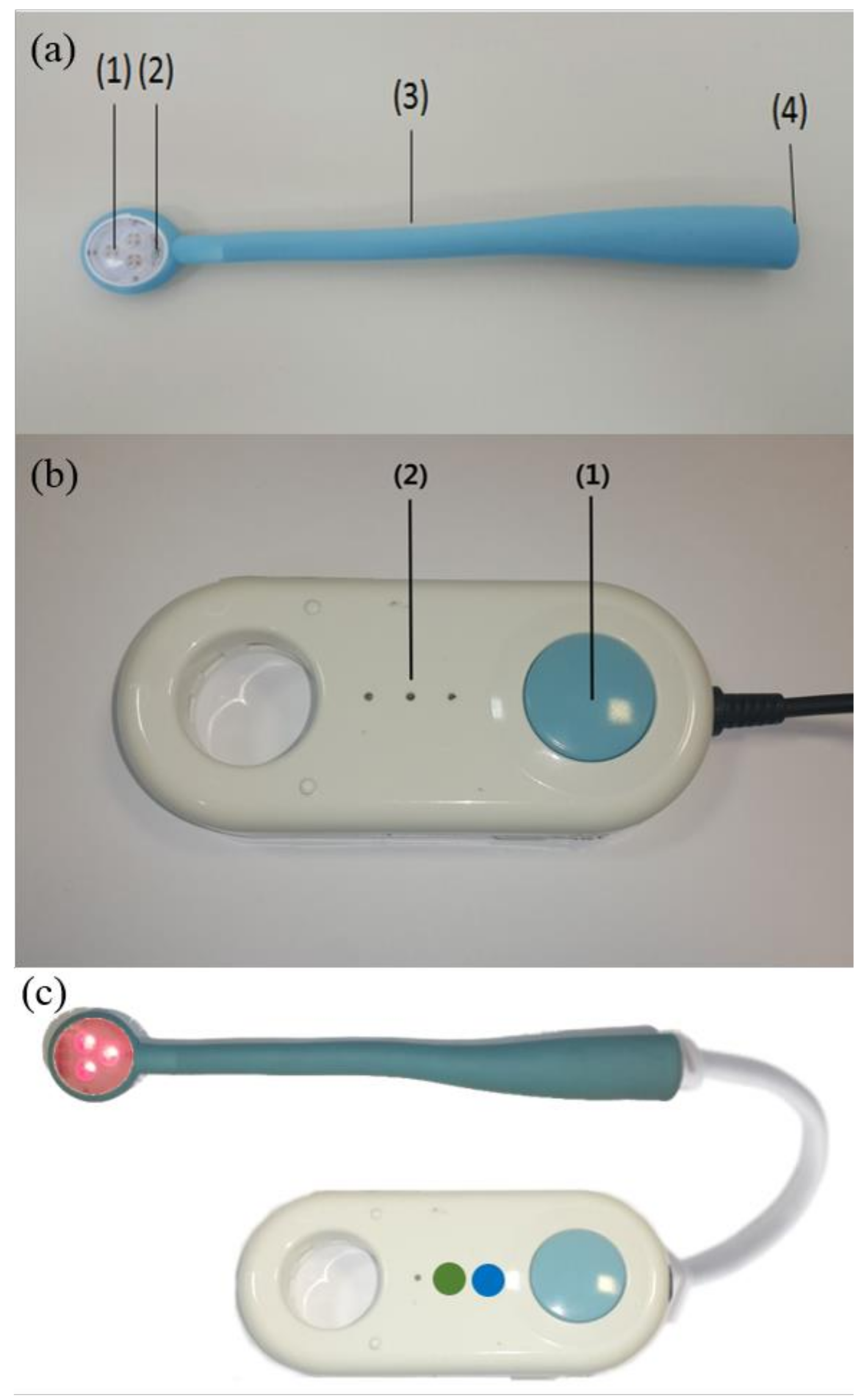
2.2. The New Handheld PBMT Device

The light source was PetalB (Ptech Corp., Pyeongtaek, Korea), which comprised four different wavelengths, AIGaInP (670 nm), AIGaAs (780 nm), GaAs (830 nm), and InGaAs (910 nm), each consisting of three laser diodes. We selected various wavelengths that were effective for OM by referring to previous studies [33]. Four wavelengths were selected, considering the treatment effect and reproducibility among the candidate wavelengths, and the light source was constructed using these wavelengths. This device performs cross-output with a 625-Hz pulse with 200 μs-on and 1400 μs-off using the four wavelengths mentioned above. Since the applicable semiconductor laser chip was selected and used according to the power level of each laser wavelength, different average powers were used according to each laser radiation source (Table 2). The laser beams from diodes were dispersed through lenses to create divergent beams. PetalB is designed in the form of a dental mirror that can be easily inserted into the oral cavity; by installing a laser diode instead of a mirror, close-up irradiation is possible even in inflamed areas of the corners of the mouth. This device is a prototype constructed by Ptech Corp. and is not yet commercially available (Figure 1).

2.3. Treatment Protocol

Patients were treated 3 or 4 days/week from the start of moderate-to-severe OM (WHO grade II, III) until the end of CRT. Laser therapy was administered through a non-contact modality (in which the laser irradiation part was covered with a disposable vinyl cover and the distance from the oral mucosa was kept close to approximately 0.2–0.5 cm) over the entire oral cavity, both in ulcerated and erythematous OM lesions, and in areas free of clinical OM signs. Eleven points in the oral cavity were irradiated: the upper lip mucosa, lower lip mucosa, upper gingiva, lower gingiva, right side of the tongue, left side of the tongue, right buccal mucosa, left buccal mucosa, hard palate, soft palate, and mouth floor. Laser therapy was applied at each point for 20 s; the protocol was repeated five times at 2–3 min intervals, and the total duration was 25–30 min. This treatment protocol was based on previous studies using PBMT in OM patients with cancer [33,37,53]. Special protective glasses were used to ensure patient and operator safety. All patients received the same instructions regarding routine oral hygiene care and standard topical/analgesic treatments for OM during treatment. Post-treatment follow-up assessments were conducted 2 and 4 weeks later. Laser therapy parameters measured using the power meter are listed in Table 2.

2.4. Clinical Efficacy and Patients’ Quality of Life

The participants were carefully assessed weekly from baseline and followed up until 4 weeks after treatment. Standardized digital photographs of the oral cavity were taken during each visit using identical positions and camera settings to ensure the reliability of the evaluation. Efficacy was assessed by evaluating global severity according to the WHO grading of mucositis scale. All adverse effects, including mucosal dryness, erythema, pruritus, and desquamation, were recorded in detail throughout the study. Patients’ quality of life was evaluated weekly using EORTC QLQ-C30 (version 3.0), and follow-ups were performed until 4 weeks after the final treatment. The EORTC QLQ-C30 comprises 30 questions, both multi-item scales and single-item measures, including five functional scales (physical functioning [PF2], role functioning [RF2], emotional functioning [EF], cognitive functioning [CF], and social functioning [SF]), three symptom scales (fatigue [FA], nausea and vomiting [NV], and pain [PA]), a global health status/quality of life scale, and six single items (dyspnea [DY], insomnia [SL], appetite loss [AP], constipation [CO], diarrhea [DI], and financial difficulties [FI]).

2.5. Metagenomics Analysis Based on 16S rRNA Gene for the Salivary Microbiome

Saliva was collected from the patients using a dedicated kit for saliva microbiome analysis (OMNIgeneORAL, OM501, DNAgenotek, Kanata, ON, Canada). The collected samples were stored in a freezer and quickly delivered to Soonchunhyang University Probiotics Microbiome Convergence Center (PMC) in a frozen state in batches.
DNA extraction from saliva samples was performed using the phenol:chloroform:isoamyl (PCI) method. The sample was centrifuged at 4000 rpm for 30 min, the supernatant was removed to obtain a pellet, and 360 μL of ATL solution was added and mixed. Afterward, 40 μL proteinase K solution (800 U/mL) was added and incubated at 56 ℃ for 3–4 h after mixing by pipetting, followed by the addition of 400 μL AL solution and incubation at 70 °C for 10 min. It was then transferred to a tube containing beads of 0.1 mm diameter (Lysing Matrix B, MP Biomedicals, Irvine, CA, USA) and subjected to physical disruption. An equal volume of 25:24:1 ratio of PCI alcohol solution was mixed and centrifuged at 12,000 rpm for 7 min at room temperature, and the upper aqueous phase (200–500 μL) was transferred to a new tube. Freeze-cold/pre-chilled 100% ethanol corresponding to 2.5 times and 3 M sodium acetate corresponding to 1/10 were mixed. After being placed at −80 °C for 1 h overnight, it was centrifuged at 15,000 rpm for 20 min at 4 °C, and the resulting pellet was washed with 500 μL of 70% ethanol. Finally, it was centrifuged again at 15,000 rpm for 5–7 min at 4 °C, and the pellet was dried for 5–10 min before resuspending in 100 μL of PCR-grade water.
The entire process of 16S gene-based metagenomic analysis, including metagenomic library formation and sequencing and data analysis, was performed according to previous reports conducted in our laboratory [54,55]. Briefly, the extracted DNA was first amplified in the V4 region of the 16S rRNA gene using primers containing overhang sequences that were compatible with the Illumina Nextera XT index. The primer sequences used were as follows: 515F(5′-TCGTCGGCAGCGTCAGATGTGTATAAGAGACAG-GTGCCAGCMGCCGCGGTAA-3′)/806R(5′GTCTCGTGGGCTCGGAGATGTGTATAAGAGACAG-GGACTACHVGGGTWTCTAAT-3′). Afterward, metagenomic libraries were prepared using the Nextera XT DNA Library Prep Kit (Illumina, San Diego, CA, USA). All PCR reactions were performed using 2 × KAPA HiFi HotStart ReadyMix (Kapa Biosystems, Wilmington, MA, USA), and PCR cleanup was performed using AMPure XP beads (Beckman Coulter, High Wycombe, UK) after each step. Samples were finally diluted from 1 nM to 50 pM in 10 mM Tris (pH 8.5), mixed with 10% PhiX, loaded onto iSeq-100 reagent cartridges (Illumina, San Diego, CA, USA), and sequenced on the iSeq-100 (Illumina, San Diego, CA, USA).

2.6. Statistical Analysis

The treatment effects and results of the EORTC QLQ-C30 were compared with the baseline scores at each follow-up visit using paired t-tests. Data were analyzed using SPSS version 26.0; IBM Corp., Armonk, NY, USA). Statistical significance was set at p < 0.05.

3. Results

3.1. Efficacy of PBMT in the Study Population

The study included 21 patients with head and neck cancer and moderate-to-severe OM. The baseline characteristics of the patients are listed in Table 3. The patients were Asians (33.3% females and 66.7% males) with a median age of 63.9 (47–93) years. All the patients had OM grade II or III OM before the start of PBMT. The mean value of OM grade decreased significantly from 2.63 ± 0.50 before PBMT to 2.13 ± 0.89, 1.31 ± 0.60 and 0.75 ± 0.58 after treatment, 2 weeks after treatment, and 4 weeks after treatment, respectively (p < 0.05, p < 0.001, and p < 0.001), as shown in Figure 2. The distribution of OM grades before and after PBMT is presented in Table 4. A significant decrease in OM severity was observed after PBMT. The proportion of patients with OM grade 2 (moderate) was 37.5% at baseline, which decreased to 4.8% at the final visit. Additionally, the proportion of patients with OM grade 3 (severe) was 62.5% at baseline; however, no patients with OM grade 3 were observed at the final visit. Clinical pictures of the two patients with marked improvement are shown in Figure 3 and Figure 4.

3.2. Evaluation of Health-Related Quality of Life (HRQoL) by EORTC QLQ-C30 Questionnaires

In the EORTC QLQ-C30, compared to baseline, we observed that patients had significantly lower scores after PBMT on all five functional scales: PF2 (p < 0.001), RF (p < 0.001), EF (p < 0.001), CF (RF (p = 0.005), and SF (p < 0.001). Additionally, patients had significantly lower scores after PBMT than at baseline on all three symptom scales: FA (p < 0.001), NV (p < 0.001), and PA (p < 0.001). Among the single items, patients had significantly lower DY (p < 0.001), SL (p = 0.001), and AP (p < 0.001) scores after PBMT. In addition, CO, DO, and FI scores were reduced after PBMT, although the reduction was not significant (p = 0.806, 0.270, and 0.136, respectively). After PBMT, the general global health status significantly increased (p < 0.001) compared to baseline, indicating improved HRQoL (Table 5).

3.3. Safety and Patient Compliance

Of the 21 patients enrolled, five dropped out; the reasons for difficulty in visiting the hospital were as follows: two patients found it difficult to visit because of too many treatment sessions, two lived too far from the hospital, and one reported that all OM lesions had healed mid-treatment. No severe side effects or side effects (e.g., mucosal dryness, erythema, pruritus, or desquamation) were reported.

3.4. Effects of PBMT Device on the Microbiome Composition in the Saliva of Patients

In the permutational multivariate analysis of variance (PERMANOVA), no significant changes in the bacterial community were identified on the species and genus criteria for samples before and after PBMT (Table 6). In addition, principal coordinate analysis (PCoA) and the unweighted pair group method with arithmetic mean (UPGMA) analyses did not show a clear distinction in the bacterial community by PBMT (Figure 5). For alpha diversity, no significant differences in species richness and diversity for the microbiome were detected in saliva samples before and after PBMT (Figure 6). Figure 7 presents the average taxonomic composition of the salivary microbial communities. Taxa with relative abundances greater than 1% in all ranks showed no significant changes with PBMT. Detailed values are presented in Table 7. Taken together, these results suggest that PBMT using this device does not have a detrimental effect on the oral microbiome.

4. Discussion

OM is a common, painful, and debilitating complication of cancer therapies. OM occurs at varying frequencies during CT and/or RT, mainly depending on the type of treatment regimen and degree of toxicity. Representative anticancer drugs that cause OM easily include high-dose methotrexate, 5-fluorouracil, and busulfan. RT, especially RT targeting the head and neck regions, is another risk factor for severe OM [56]. Approximately 90% of head and neck cancer patients receiving RT developed OM [57,58], leading to huge medical expenditure. Severe OM is an unmet medical need in head and neck cancer patients undergoing CRT because it significantly affects quality of life and interferes with cancer therapy; however, no effective preventive strategy is currently available [59]. Previous studies on cancer therapy-induced OM reported good results with the use of PBMT in OM prevention [33].
This study was conducted to explore the effectiveness of a novel handheld PBMT device for treating CRT-induced OM in patients with head and neck cancer. Overall, our results showed that this device was effective in treating CRT-induced OM with excellent patient compliance. After PBMT, we observed a significant decrease in the mean WHO OM grade, and an additional significant decrease was observed at the final 4-week follow-up. In addition, no difficulties were encountered in administering PBMT and no side effects were documented. Overall, the patients enrolled in this study had strong compliance and subjective satisfaction with the treatment device. If further large-scale studies report similar effectiveness and safety, this novel handheld PBMT device may become a reliable and safe alternative for the treatment of CRT-induced OM in patients with head and neck cancer. Similar to our results, no serious side effects have been reported to date in previous clinical studies applying PBMT for the management of cancer therapy-induced OM [60,61]. Despite the various mechanisms that alter its biological activity, the device is considered safe when used appropriately.
In addition, the patients’ quality of life as measured using the EORTC QLQ-C30 showed an overall improvement after PBMT. The rate of OM grade II or lower after PBMT was significantly higher than that before treatment, which is thought to have affected the patient’s perceived quality of life. Patients with OM grade ≤ II are generally able to eat adequate amounts of food and maintain their nutritional status. From this perspective, lowering the OM grade to II or lower has a positive impact on patients’ quality of life through the restoration of feeding capacity, a strong indicator of patient well-being. Malnutrition can also affect the response and adherence to cancer treatment by increasing the risk of toxicity and infection [62]. In this study, the participants visited the hospital and received PBMT; however, in the future, this handheld device may be used for treatment at home instead of the patient visiting the clinic regularly, which requires less time and effort, potentially leading to excellent compliance.
Factors such as wavelength, energy density, and irradiation frequency can influence the cellular mechanisms of PBMT [63]. The wavelength of PBMT is an important factor that affects cellular response. Red to near-infrared light in the range of 600–1070 nm is known to have the greatest effect on cellular and molecular responses, including cell proliferation, metabolism, and angiogenesis. This phenomenon may be due to the absorption or interference of light beyond this range. It is thought that shorter wavelengths can be absorbed by hemoglobin or melanin, whereas longer wavelengths penetrate more deeply but can be absorbed by water; thus, only a specific wavelength has effective biostimulatory effects [64,65,66]. Ma et al. reported that PBMT using a dual wavelength of 635 nm and 830 nm increased fibroblast proliferation and collagen synthesis, whereas PBMT using a single wavelength of 635 nm had no significant effect [67]. Recently, another in vitro and in vivo study reported that a 660 nm wavelength increased ROS production, and a 970 nm wavelength showed antioxidant activity. Interestingly, a significant reduction in ROS levels was detected in cells exposed to the 800 nm wavelength or a combination of the three different wavelengths (660 nm, 800 nm, and 970 nm). These findings suggest that multi-wavelength PBMT may be clinically useful by efficiently reaching various tissue depths and exploiting the diverse properties of each wavelength [68]. Thus, we decided to use laser diodes with four different wavelengths (670 nm, 780 nm, 830 nm, and 910 nm) in the present study and obtained promising results. However, future in vitro, in vivo, and clinical studies are required to confirm the rationale of our protocol.
There was no significant difference in oral microbiome composition before and after PBMT. Sufficient microbiome recovery was difficult because PBMT was applied after severe dysbiosis had already occurred via CRT. Nevertheless, the results of this study confirm that PBMT maintains homeostasis without adversely affecting the oral microbiome. However, this study is limited in that changes at the taxonomic level were investigated, and considering that the near-infrared energy of PBMT can affect the metabolism of bacteria [30,69], a holistic understanding based on the analysis of the entire genome of the microbiome is required. Some studies showed that the visible and near-infrared wavelengths of PBMT may affect oral bacterial growth and salivary gland activity by modulating the bacterial cell cycle through interactions on photoacceptors such as cytochrome, flavins and voltage-dependent calcium channels [69,70]. In the future, a well-designed large-scale study is needed to analyze the effects of PBMT on the oral microbiome over a long period, including before the start of cancer treatment and a control group. In addition, a detailed analysis is necessary depending on the type and duration of anticancer drugs, whether RT was used, and the severity of OM. In addition, in-depth research is needed on the mechanism by which PBMT prevents the destruction of microorganisms during cancer treatment and the relationship between the improvement in OM and equilibrium of the oral microbiome.
This study had some limitations. Although our results are promising, an optimal irradiation fluence and treatment regimen (including the distance between the device and oral mucosa) should be determined in future studies. In addition, this study had a significant limitation of not having a control group. Treatment with this device is a non-contact modality, and the distance to the oral mucosa may not be constant, which may affect the amount of energy reaching the tissue; therefore, there is a possibility of bias. In addition, we did not evaluate the oral hygiene status. Many studies have reported that the maintenance of good oral health reduces OM severity, but also helps in its resolution [71]. Other studies have reported that compromised immune status, poor oral hygiene, and pre-existing oral damage such as tooth decay and plaque are associated with a very severe OM [72]. The MASCC/ISOO guidelines recommend the use of a standardized oral care protocol, including brushing with a soft toothbrush, flossing, and non-medicated rinses [73]. Furthermore, the possible preventive role of PBMT was not investigated in the present study. An effective role of PBMT in reducing the incidence and severity of OM when administered before symptom onset has been reported [74]. Further studies using larger randomized controlled trials are required to obtain better evidence for the clinical applications of this novel PBMT device. As the molecular mechanisms underlying the advantages and clinical efficacy of the combined irradiation of the four wavelengths used in this study have not been fully explored, additional follow-up studies are required.

5. Conclusions

OM is a common side effect associated with conventional cancer therapy, and when it is severe, it not only has a major impact on patients’ quality of life, but also causes other side effects such as secondary infections. However, no standard treatment has been suggested to date. Therefore, alternative treatment options for OM are required. Thus, this study was conducted to explore the effectiveness of a novel handheld PBMT device for treating CRT-induced OM in patients with head and neck cancer. In conclusion, our results showed that this new portable device is effective in treating CRT-induced OM with excellent patient compliance. After PBMT, a significant decrease in the mean WHO OM grade was observed. In addition, we observed a significant improvement in HRQoL after PBMT compared with baseline in the EORTC QLQ-C30 results. PBMT using this novel device could be a supportive therapeutic modality for treating OM in patients with head and neck cancer. Further mechanistic and large-scale randomized controlled studies on cancer therapy-associated OM should be carried out to determine its effectiveness and safety and investigate the exact mechanisms.

Supplementary Materials

The following supporting information can be downloaded at: https://www.mdpi.com/article/10.3390/photonics10030241/s1, Table S1: 2013 CARE Checklist of information to include when writing a case report.

Author Contributions

Conceptualization, I.-Y.J. and H.-K.B.; methodology, M.-J.B. and J.-H.P.; software, N.-H.H.; validation, S.-C.L. and Y.-K.W.; formal analysis, N.-H.H.; investigation, H.S., S.K. and P.L.; resources, S.J. and N.-G.Y.; data curation, H.-Y.S. and J.-Y.K.; writing—original draft preparation, Y.-S.E. and J.-Y.K.; writing—review and editing, I.-Y.J., H.-K.B. and S.-H.L.; visualization, I.-Y.J.; supervision, J.-E.K.; project administration, J.-E.K.; funding acquisition, J.-E.K. All authors have read and agreed to the published version of the manuscript.

Funding

This research was supported by the Soonchunhyang University Research Fund, National Research Foundation of Korea (grant no. NRF-2021R1F1A1052669), and the Korean Medical Device Development Fund grant funded by the Korean government (Ministry of Science and ICT, Ministry of Trade Industry and Energy, Ministry of Health and Welfare, and Ministry of Food and Drug Safety) [Project No. RS-2020-KD000281].

Institutional Review Board Statement

The study was conducted in accordance with the Declaration of Helsinki, and approved by the Institutional Review Board of Soonchunhyang University Cheonan Hospital (IRB No. 2020-06-032-028) and Soonchunhyang University Seoul Hospital (IRB No. 2020-09-014).

Informed Consent Statement

Informed consent was obtained from all the participants involved in the study. Written informed consent was obtained from the patients for the publication of this paper.

Data Availability Statement

The data used to support the results of this study are available from the corresponding author upon request.

Conflicts of Interest

Ptech Corp. developed and provided the photobiomodulation devices used in the study. The funders (Soonchunhyang University Research Fund, National Research Foundation of Korea, and the Korean Medical Device Development Fund grant funded by Korean government) had no role in the study design; collection, analyses, or interpretation of data; writing of the manuscript; or decision to publish the results.

References

  1. Legouté, F.; Bensadoun, R.-J.; Seegers, V.; Pointreau, Y.; Caron, D.; Lang, P.; Prévost, A.; Martin, L.; Schick, U.; Morvant, B. Low-level laser therapy in treatment of chemoradiotherapy-induced mucositis in head and neck cancer: Results of a randomised, triple blind, multicentre phase III trial. Radiat. Oncol. 2019, 14, 83. [Google Scholar] [CrossRef] [PubMed]
  2. Bolton, L. Managing Oral Mucositis in Patients with Cancer. Wounds 2021, 33, 136–138. [Google Scholar] [CrossRef] [PubMed]
  3. Cronshaw, M.; Parker, S.; Anagnostaki, E.; Mylona, V.; Lynch, E.; Grootveld, M. Photobiomodulation and oral mucositis: A systematic review. Dent. J. 2020, 8, 87. [Google Scholar] [CrossRef] [PubMed]
  4. Anderson, C.M.; Lee, C.M.; Saunders, D.P.; Curtis, A.; Dunlap, N.; Nangia, C.; Lee, A.S.; Gordon, S.M.; Kovoor, P.; Arevalo-Araujo, R.; et al. Phase IIb, Randomized, Double-Blind Trial of GC4419 Versus Placebo to Reduce Severe Oral Mucositis Due to Concurrent Radiotherapy and Cisplatin for Head and Neck Cancer. J. Clin. Oncol. 2019, 37, 3256–3265. [Google Scholar] [CrossRef] [PubMed]
  5. Henke, M.; Alfonsi, M.; Foa, P.; Giralt, J.; Bardet, E.; Cerezo, L.; Salzwimmer, M.; Lizambri, R.; Emmerson, L.; Chen, M.G.; et al. Palifermin decreases severe oral mucositis of patients undergoing postoperative radiochemotherapy for head and neck cancer: A randomized, placebo-controlled trial. J. Clin. Oncol. 2011, 29, 2815–2820. [Google Scholar] [CrossRef] [PubMed]
  6. Riley, P.; Glenny, A.M.; Worthington, H.V.; Littlewood, A.; Mauleffinch, L.M.F.; Clarkson, J.E.; McCabe, M.G. Interventions for preventing oral mucositis in patients with cancer receiving treatment: Cytokines and growth factors. Cochrane Database Syst. Rev. 2017, 11, CD011990. [Google Scholar] [CrossRef]
  7. Oton-Leite, A.F.; Silva, G.B.L.; Morais, M.O.; Silva, T.A.; Leles, C.R.; Valadares, M.C.; Pinezi, J.C.D.; Batista, A.C.; Mendonça, E.F. Effect of low-level laser therapy on chemoradiotherapy-induced oral mucositis and salivary inflammatory mediators in head and neck cancer patients. Lasers Surg. Med. 2015, 47, 296–305. [Google Scholar] [CrossRef]
  8. Bowen, J.; Al-Dasooqi, N.; Bossi, P.; Wardill, H.; Van Sebille, Y.; Al-Azri, A.; Bateman, E.; Correa, M.E.; Raber-Durlacher, J.; Kandwal, A.; et al. The pathogenesis of mucositis: Updated perspectives and emerging targets. Support. Care Cancer 2019, 27, 4023–4033. [Google Scholar] [CrossRef]
  9. Parker, S.; Cronshaw, M.; Grootveld, M. Photobiomodulation Delivery Parameters in Dentistry: An Evidence-Based Approach. Photobiomodul. Photomed. Laser. Surg. 2022, 40, 42–50. [Google Scholar] [CrossRef]
  10. Caillot, E.; Denis, F. Radio-induced oral and pharyngeal mucositis: Management updates. Cancer Radiother. 2012, 16, 358–363. [Google Scholar] [CrossRef]
  11. Allison, R.R.; Ambrad, A.A.; Arshoun, Y.; Carmel, R.J.; Ciuba, D.F.; Feldman, E.; Finkelstein, S.E.; Gandhavadi, R.; Heron, D.E.; Lane, S.C.; et al. Multi-institutional, randomized, double-blind, placebo-controlled trial to assess the efficacy of a mucoadhesive hydrogel (MuGard) in mitigating oral mucositis symptoms in patients being treated with chemoradiation therapy for cancers of the head and neck. Cancer 2014, 120, 1433–1440. [Google Scholar] [CrossRef] [PubMed]
  12. Barber, C.; Powell, R.; Ellis, A.; Hewett, J. Comparing pain control and ability to eat and drink with standard therapy vs Gelclair: A preliminary, double centre, randomised controlled trial on patients with radiotherapy-induced oral mucositis. Support Care Cancer 2007, 15, 427–440. [Google Scholar] [CrossRef] [PubMed]
  13. Leenstra, J.L.; Miller, R.C.; Qin, R.; Martenson, J.A.; Dornfeld, K.J.; Bearden, J.D.; Puri, D.R.; Stella, P.J.; Mazurczak, M.A.; Klish, M.D.; et al. Doxepin rinse versus placebo in the treatment of acute oral mucositis pain in patients receiving head and neck radiotherapy with or without chemotherapy: A phase III, randomized, double-blind trial (NCCTG-N09C6 [Alliance]). J. Clin. Oncol. 2014, 32, 1571–1577. [Google Scholar] [CrossRef] [PubMed]
  14. Rao, N.G.; Trotti, A.; Kim, J.; Schell, M.J.; Zhao, X.; Amdur, R.J.; Brizel, D.M.; Chambers, M.S.; Caudell, J.J.; Miyamoto, C.; et al. Phase II multicenter trial of Caphosol for the reduction of mucositis in patients receiving radiation therapy for head and neck cancer. Oral Oncol. 2014, 50, 765–769. [Google Scholar] [CrossRef]
  15. Sarvizadeh, M.; Hemati, S.; Meidani, M.; Ashouri, M.; Roayaei, M.; Shahsanai, A. Morphine mouthwash for the management of oral mucositis in patients with head and neck cancer. Adv. Biomed. Res. 2015, 4, 44. [Google Scholar] [CrossRef]
  16. Wong, K.H.; Kuciejewska, A.; Sharabiani, M.T.A.; Ng-Cheng-Hin, B.; Hoy, S.; Hurley, T.; Rydon, J.; Grove, L.; Santos, A.; Ryugenji, M.; et al. A randomised controlled trial of Caphosol mouthwash in management of radiation-induced mucositis in head and neck cancer. Radiother. Oncol. 2017, 122, 207–211. [Google Scholar] [CrossRef]
  17. Sio, T.T.; Le-Rademacher, J.G.; Leenstra, J.L.; Loprinzi, C.L.; Rine, G.; Curtis, A.; Singh, A.K.; Martenson, J.A., Jr.; Novotny, P.J.; Tan, A.D.; et al. Effect of Doxepin Mouthwash or Diphenhydramine-Lidocaine-Antacid Mouthwash vs Placebo on Radiotherapy-Related Oral Mucositis Pain: The Alliance A221304 Randomized Clinical Trial. JAMA 2019, 321, 1481–1490. [Google Scholar] [CrossRef]
  18. Damani, A.; Salins, N.; Ghoshal, A.; Muckaden, M. Specialist Pediatric Palliative Care Prescribing Practices: A Large 5-year Retrospective Audit. Indian J. Palliat. Care 2016, 22, 274–281. [Google Scholar] [CrossRef]
  19. Hu, W.; Chen, L.; Wang, C.; Guo, L.; Hua, X.; Cai, Y.; Fan, Y. Standardized nursing and therapeutic effect of oxycontin on oral mucosal pain in nasopharyngeal carcinoma patients. J. Cancer Res. Ther. 2018, 14, 1594–1599. [Google Scholar] [CrossRef]
  20. Kim, J.W. Clinical Trial of Nonthermal 633 nm Omnilux LED Array for Renewal of Photoaging: Clinical Surface Profilometric Results. J. Korean Soc. Laser Med. 2005, 9, 69–76. [Google Scholar]
  21. Nestor, M.; Andriessen, A.; Berman, B.; Katz, B.E.; Gilbert, D.; Goldberg, D.J.; Gold, M.H.; Kirsner, R.S.; Lorenc, P.Z. Photobiomodulation with non-thermal lasers: Mechanisms of action and therapeutic uses in dermatology and aesthetic medicine. J. Cosmet. Laser Ther. 2017, 19, 190–198. [Google Scholar] [CrossRef] [PubMed]
  22. Schubert, M.M.; Eduardo, F.P.; Guthrie, K.A.; Franquin, J.-C.; Bensadoun, R.-J.J.; Migliorati, C.A.; Lloid, C.M.E.; Eduardo, C.P.; Walter Filho, N.; Marques, M.M. A phase III randomized double-blind placebo-controlled clinical trial to determine the efficacy of low level laser therapy for the prevention of oral mucositis in patients undergoing hematopoietic cell transplantation. Support Care Cancer 2007, 15, 1145–1154. [Google Scholar] [CrossRef] [PubMed]
  23. De Freitas, L.F.; Hamblin, M.R. Proposed Mechanisms of Photobiomodulation or Low-Level Light Therapy. IEEE J. Sel. Top. Quantum Electron. 2016, 22, 348–364. [Google Scholar] [CrossRef]
  24. Hamblin, M.R. Mechanisms and applications of the anti-inflammatory effects of photobiomodulation. AIMS Biophys. 2017, 4, 337–361. [Google Scholar] [CrossRef] [PubMed]
  25. Rola, P.; Włodarczak, S.; Lesiak, M.; Doroszko, A.; Włodarczak, A. Changes in Cell Biology under the Influence of Low-Level Laser Therapy. Photonics 2022, 9, 502. [Google Scholar] [CrossRef]
  26. Peng, H.; Chen, B.B.; Chen, L.; Chen, Y.P.; Liu, X.; Tang, L.L.; Mao, Y.P.; Li, W.F.; Zhang, Y.; Lin, A.H.; et al. A network meta-analysis in comparing prophylactic treatments of radiotherapy-induced oral mucositis for patients with head and neck cancers receiving radiotherapy. Oral Oncol. 2017, 75, 89–94. [Google Scholar] [CrossRef] [PubMed]
  27. Lalla, R.V.; Brennan, M.T.; Gordon, S.M.; Sonis, S.T.; Rosenthal, D.I.; Keefe, D.M. Oral Mucositis Due to High-Dose Chemotherapy and/or Head and Neck Radiation Therapy. J. Natl. Cancer Inst. Monogr. 2019, 2019(53), lgz011. [Google Scholar] [CrossRef]
  28. Sardo, A.V.N.; Andrade, M.F.; Figueiredo, A.; Rosin, F.C.P.; Corrêa, L.; Zezell, D.M. Does Photobiomodulation Affects CK10 and CK14 in Oral Mucositis Radioinduced Repair? Int. J. Mol. Sci. 2022, 23, 15611. [Google Scholar] [CrossRef]
  29. Andrade, M.F.; Sardo, A.V.N.; Benetti, C.; Sicchieri, L.B.; Corrêa, L.; Zezell, D.M. Comparison of Two Light Wavelengths (&lambda; = 660 nm and &lambda; = 780 nm) in the Repair Process of Oral Mucositis Induced by Ionizing Radiation: Clinical and Microscopic Evaluations in Rats. Photonics 2023, 10, 16. [Google Scholar]
  30. Avci, P.; Gupta, A.; Sadasivam, M.; Vecchio, D.; Pam, Z.; Pam, N.; Hamblin, M.R. Low-level laser (light) therapy (LLLT) in skin: Stimulating, healing, restoring. Semin. Cutan. Med. Surg. 2013, 32, 41–52. [Google Scholar]
  31. Zanin, T.; Zanin, F.; Carvalhosa, A.A.; de Souza Castro, P.H.; Pacheco, M.T.; Zanin, I.C.J.; Brugnera, A. Use of 660-nm diode laser in the prevention and treatment of human oral mucositis induced by radiotherapy and chemotherapy. Photomed. Laser Surg. 2010, 28, 233–237. [Google Scholar] [CrossRef] [PubMed]
  32. Rubenstein, E.B.; Peterson, D.E.; Schubert, M.; Keefe, D.; McGuire, D.; Epstein, J.; Elting, L.S.; Fox, P.C.; Cooksley, C.; Sonis, S.T. Clinical practice guidelines for the prevention and treatment of cancer therapy-induced oral and gastrointestinal mucositis. Cancer 2004, 100, 2026–2046. [Google Scholar] [CrossRef] [PubMed]
  33. Zadik, Y.; Arany, P.R.; Fregnani, E.R.; Bossi, P.; Antunes, H.S.; Bensadoun, R.-J.; Gueiros, L.A.; Majorana, A.; Nair, R.G.; Ranna, V. Systematic review of photobiomodulation for the management of oral mucositis in cancer patients and clinical practice guidelines. Support. Care Cancer 2019, 27, 3969–3983. [Google Scholar] [CrossRef] [PubMed]
  34. Elad, S.; Cheng, K.K.F.; Lalla, R.V.; Yarom, N.; Hong, C.; Logan, R.M.; Bowen, J.; Gibson, R.; Saunders, D.P.; Zadik, Y. MASCC/ISOO clinical practice guidelines for the management of mucositis secondary to cancer therapy. Cancer 2020, 126, 4423–4431. [Google Scholar] [CrossRef] [PubMed]
  35. Lee, W.J.; Lee, K.C.; Kim, M.J.; Jang, Y.H.; Lee, S.J.; Kim, D.W. Efficacy of Red or Infrared Light-Emitting Diodes in a Mouse Model of Propionibacterium acnes-Induced Inflammation. Ann. Dermatol. 2016, 28, 186–191. [Google Scholar] [CrossRef]
  36. Ramos, R.R.; Paiva, J.L.; Gomes, J.; Boer, N.P.; Godoy, J.M.P.; Batigalia, F. Photodynamic action of the red laser on Propionibacterium acnes. An. Bras. Dermatol. 2017, 92, 622–625. [Google Scholar] [CrossRef]
  37. Gobbo, M.; Verzegnassi, F.; Ronfani, L.; Zanon, D.; Melchionda, F.; Bagattoni, S.; Majorana, A.; Bardellini, E.; Mura, R.; Piras, A. Multicenter randomized, double-blind controlled trial to evaluate the efficacy of laser therapy for the treatment of severe oral mucositis induced by chemotherapy in children: laMPO RCT. Pediatr. Blood Cancer 2018, 65, e27098. [Google Scholar] [CrossRef]
  38. Ottaviani, G.; Gobbo, M.; Sturnega, M.; Martinelli, V.; Mano, M.; Zanconati, F.; Bussani, R.; Perinetti, G.; Long, C.S.; Di Lenarda, R. Effect of class IV laser therapy on chemotherapy-induced oral mucositis: A clinical and experimental study. Am. J. Pathol. 2013, 183, 1747–1757. [Google Scholar] [CrossRef]
  39. Dose, A.M. The symptom experience of mucositis, stomatitis, and xerostomia. In Seminars in Oncology Nursing; WB Saunders: Philadelphia, PA, USA, 1995; pp. 248–255. [Google Scholar]
  40. Khouri, V.Y.; Stracieri, A.B.P.L.; Rodrigues, M.C.; Moraes, D.A.d.; Pieroni, F.; Simões, B.P.; Voltarelli, J.C. Use of therapeutic laser for prevention and treatment of oral mucositis. Braz. Dent. J. 2009, 20, 215–220. [Google Scholar] [CrossRef]
  41. Helmink, B.A.; Khan, M.A.W.; Hermann, A.; Gopalakrishnan, V.; Wargo, J.A. The microbiome, cancer, and cancer therapy. Nat. Med. 2019, 25, 377–388. [Google Scholar] [CrossRef]
  42. Sheflin, A.M.; Whitney, A.K.; Weir, T.L. Cancer-promoting effects of microbial dysbiosis. Curr. Oncol. Rep. 2014, 16, 406. [Google Scholar] [CrossRef] [PubMed]
  43. La Rosa, G.R.M.; Gattuso, G.; Pedullà, E.; Rapisarda, E.; Nicolosi, D.; Salmeri, M. Association of oral dysbiosis with oral cancer development. Oncol. Lett. 2020, 19, 3045–3058. [Google Scholar] [CrossRef] [PubMed]
  44. Hong, B.Y.; Sobue, T.; Choquette, L.; Dupuy, A.K.; Thompson, A.; Burleson, J.A.; Salner, A.L.; Schauer, P.K.; Joshi, P.; Fox, E.; et al. Chemotherapy-induced oral mucositis is associated with detrimental bacterial dysbiosis. Microbiome 2019, 7, 66. [Google Scholar] [CrossRef] [PubMed]
  45. Perales-Puchalt, A.; Perez-Sanz, J.; Payne, K.K.; Svoronos, N.; Allegrezza, M.J.; Chaurio, R.A.; Anadon, C.; Calmette, J.; Biswas, S.; Mine, J.A.; et al. Frontline Science: Microbiota reconstitution restores intestinal integrity after cisplatin therapy. J. Leukoc. Biol. 2018, 103, 799–805. [Google Scholar] [CrossRef] [PubMed]
  46. Zagury-Orly, I.; Khaouam, N.; Noujaim, J.; Desrosiers, M.Y.; Maniakas, A. The Effect of Radiation and Chemoradiation Therapy on the Head and Neck Mucosal Microbiome: A Review. Front. Oncol. 2021, 11, 784457. [Google Scholar] [CrossRef]
  47. Zanotta, N.; Ottaviani, G.; Campisciano, G.; Poropat, A.; Bovenzi, M.; Rupel, K.; Gobbo, M.; Comar, M.; Di Lenarda, R.; Biasotto, M.; et al. Photobiomodulation modulates inflammation and oral microbiome: A pilot study. Biomarkers 2020, 25, 677–684. [Google Scholar] [CrossRef]
  48. Tilg, H.; Kaser, A. Gut microbiome, obesity, and metabolic dysfunction. J. Clin. Investig. 2011, 121, 2126–2132. [Google Scholar] [CrossRef]
  49. Bicknell, B.; Liebert, A.; Johnstone, D.; Kiat, H. Photobiomodulation of the microbiome: Implications for metabolic and inflammatory diseases. Lasers Med. Sci. 2019, 34, 317–327. [Google Scholar] [CrossRef]
  50. Nguyen, T.L.; Vieira-Silva, S.; Liston, A.; Raes, J. How informative is the mouse for human gut microbiota research? Dis. Model. Mech. 2015, 8, 1–16. [Google Scholar] [CrossRef]
  51. Gold, M.H.; Sensing, W.; Biron, J.A. Clinical efficacy of home-use blue-light therapy for mild-to moderate acne. J. Cosmet. Laser Ther. 2011, 13, 308–314. [Google Scholar] [CrossRef]
  52. Ash, C.; Harrison, A.; Drew, S.; Whittall, R. A randomized controlled study for the treatment of acne vulgaris using high-intensity 414 nm solid state diode arrays. J. Cosmet. Laser Ther. 2015, 17, 170–176. [Google Scholar] [CrossRef] [PubMed]
  53. Gautam, A.P.; Fernandes, D.J.; Vidyasagar, M.S.; Maiya, A.G.; Guddattu, V. Low level laser therapy against radiation induced oral mucositis in elderly head and neck cancer patients-a randomized placebo controlled trial. J. Photochem. Photobiol. B 2015, 144, 51–56. [Google Scholar] [CrossRef] [PubMed]
  54. Kim, S.; Seo, H.; Rahim, M.A.; Lee, S.; Kim, Y.S.; Song, H.Y. Changes in the Microbiome of Vaginal Fluid after Menopause in Korean Women. J. Microbiol. Biotechnol. 2021, 31, 1490–1500. [Google Scholar] [CrossRef] [PubMed]
  55. Ul-Haq, A.; Lee, K.A.; Seo, H.; Kim, S.; Jo, S.; Ko, K.M.; Moon, S.J.; Kim, Y.S.; Choi, J.R.; Song, H.Y.; et al. Characteristic alterations of gut microbiota in uncontrolled gout. J. Microbiol. 2022, 60, 1178–1190. [Google Scholar] [CrossRef]
  56. Elad, S.; Yarom, N.; Zadik, Y.; Kuten-Shorrer, M.; Sonis, S.T. The broadening scope of oral mucositis and oral ulcerative mucosal toxicities of anticancer therapies. CA Cancer J. Clin. 2022, 72, 57–77. [Google Scholar] [CrossRef]
  57. Lima, A.G.; Antequera, R.; Peres, M.P.; Snitcosky, I.M.; Federico, M.H.; Villar, R.C. Efficacy of low-level laser therapy and aluminum hydroxide in patients with chemotherapy and radiotherapy-induced oral mucositis. Braz. Dent. J. 2010, 21, 186–192. [Google Scholar] [CrossRef]
  58. Elting, L.S.; Cooksley, C.D.; Chambers, M.S.; Garden, A.S. Risk, outcomes, and costs of radiation-induced oral mucositis among patients with head-and-neck malignancies. Int. J. Radiat. Oncol. Biol. Phys. 2007, 68, 1110–1120. [Google Scholar] [CrossRef]
  59. Antunes, H.S.; Herchenhorn, D.; Small, I.A.; Araújo, C.M.; Viégas, C.M.; Cabral, E.; Rampini, M.P.; Rodrigues, P.C.; Silva, T.G.; Ferreira, E.M.; et al. Phase III trial of low-level laser therapy to prevent oral mucositis in head and neck cancer patients treated with concurrent chemoradiation. Radiother. Oncol. 2013, 109, 297–302. [Google Scholar] [CrossRef]
  60. De Pauli Paglioni, M.; Araújo, A.L.D.; Arboleda, L.P.A.; Palmier, N.R.; Fonsêca, J.M.; Gomes-Silva, W.; Madrid-Troconis, C.C.; Silveira, F.M.; Martins, M.D.; Faria, K.M.; et al. Tumor safety and side effects of photobiomodulation therapy used for prevention and management of cancer treatment toxicities. A systematic review. Oral Oncol. 2019, 93, 21–28. [Google Scholar] [CrossRef]
  61. Zecha, J.A.; Raber-Durlacher, J.E.; Nair, R.G.; Epstein, J.B.; Sonis, S.T.; Elad, S.; Hamblin, M.R.; Barasch, A.; Migliorati, C.A.; Milstein, D.M.; et al. Low level laser therapy/photobiomodulation in the management of side effects of chemoradiation therapy in head and neck cancer: Part 1: Mechanisms of action, dosimetric, and safety considerations. Support. Care Cancer 2016, 24, 2781–2792. [Google Scholar] [CrossRef]
  62. Gobbo, M.; Ottaviani, G.; Perinetti, G.; Ciriello, F.; Beorchia, A.; Giacca, M.; Di Lenarda, R.; Rupel, K.; Tirelli, G.; Zacchigna, S.; et al. Evaluation of nutritional status in head and neck radio-treated patients affected by oral mucositis: Efficacy of class IV laser therapy. Support. Care Cancer 2014, 22, 1851–1856. [Google Scholar] [CrossRef] [PubMed]
  63. Pires Oliveira, D.A.; de Oliveira, R.F.; Zangaro, R.A.; Soares, C.P. Evaluation of low-level laser therapy of osteoblastic cells. Photomed. Laser Surg. 2008, 26, 401–404. [Google Scholar] [CrossRef] [PubMed]
  64. Karu, T.I. Multiple roles of cytochrome c oxidase in mammalian cells under action of red and IR-A radiation. IUBMB Life 2010, 62, 607–610. [Google Scholar] [CrossRef] [PubMed]
  65. Sommer, A.P. Mitochondrial cytochrome c oxidase is not the primary acceptor for near infrared light-it is mitochondrial bound water: The principles of low-level light therapy. Ann. Transl. Med. 2019, 7, S13. [Google Scholar] [CrossRef]
  66. Tam, S.Y.; Tam, V.C.W.; Ramkumar, S.; Khaw, M.L.; Law, H.K.W.; Lee, S.W.Y. Review on the Cellular Mechanisms of Low-Level Laser Therapy Use in Oncology. Front. Oncol. 2020, 10, 1255. [Google Scholar] [CrossRef]
  67. Ma, H.; Yang, J.-P.; Tan, R.K.; Lee, H.-W.; Han, S.-K. Effect of Low-Level Laser Therapy on Proliferation and Collagen Synthesis of Human Fibroblasts in Vitro. J. Wound Manag. Res. 2018, 14, 1–6. [Google Scholar] [CrossRef]
  68. Rupel, K.; Zupin, L.; Colliva, A.; Kamada, A.; Poropat, A.; Ottaviani, G.; Gobbo, M.; Fanfoni, L.; Gratton, R.; Santoro, M.; et al. Photobiomodulation at Multiple Wavelengths Differentially Modulates Oxidative Stress In Vitro and In Vivo. Oxid. Med. Cell Longev. 2018, 2018, 6510159. [Google Scholar] [CrossRef]
  69. Amaroli, A.; Ravera, S.; Zekiy, A.; Benedicenti, S.; Pasquale, C. A Narrative Review on Oral and Periodontal Bacteria Microbiota Photobiomodulation, through Visible and Near-Infrared Light: From the Origins to Modern Therapies. Int. J. Mol. Sci. 2022, 23, 1372. [Google Scholar] [CrossRef]
  70. Basso, F.G.; Oliveira, C.F.; Fontana, A.; Kurachi, C.; Bagnato, V.S.; Spolidório, D.M.; Hebling, J.; de Souza Costa, C.A. In Vitro effect of low-level laser therapy on typical oral microbial biofilms. Braz Dent. J. 2011, 22, 502–510. [Google Scholar] [CrossRef]
  71. Cheng, K.K.; Molassiotis, A.; Chang, A.M.; Wai, W.C.; Cheung, S.S. Evaluation of an oral care protocol intervention in the prevention of chemotherapy-induced oral mucositis in paediatric cancer patients. Eur. J. Cancer 2001, 37, 2056–2063. [Google Scholar] [CrossRef]
  72. Lalla, R.V.; Sonis, S.T.; Peterson, D.E. Management of oral mucositis in patients who have cancer. Dent. Clin. N. Am. 2008, 52, 61–77. [Google Scholar] [CrossRef] [PubMed]
  73. McGuire, D.B.; Correa, M.E.; Johnson, J.; Wienandts, P. The role of basic oral care and good clinical practice principles in the management of oral mucositis. Support. Care Cancer 2006, 14, 541–547. [Google Scholar] [CrossRef] [PubMed]
  74. Oberoi, S.; Zamperlini-Netto, G.; Beyene, J.; Treister, N.S.; Sung, L. Effect of prophylactic low level laser therapy on oral mucositis: A systematic review and meta-analysis. PLoS ONE 2014, 9, e107418. [Google Scholar] [CrossRef] [PubMed]
Figure 1. PetalB (Ptech Corp., Pyeongtaek, Republic of Korea). (a) Description of the laser irradiation part of PBMT device. (1) Laser irradiation unit; (2) laser irradiation part cover; (3) silicone handle; (4) main body connect port. (b) Description of the main body of PBMT device. (1) Device operation button; (2) operation indicator lamp. (c) Photos of laser being irradiated by connecting the main body and the laser irradiation part.
Photonics 10 00241 g001
Figure 2. Changes in mean WHO grade of OM with time (p < 0.001).
Photonics 10 00241 g002
Figure 3. (a) 50-year-old female prior to treatments. (b) At 2 weeks after 4 weeks of PBMT treatments.
Photonics 10 00241 g003
Figure 4. (a,b) 63-year-old male prior to treatments. (c,d) At 2 weeks after 4 weeks of PBMT treatments.
Photonics 10 00241 g004
Figure 5. Beta diversity index for the salivary microbiome of cancer patients. (a) UniFrac-based PCoA (principal coordinate analysis) and (b) UPGMA (unweighted pair group method with arithmetic mean) were applied to the beta diversity analysis of the salivary microbiome.
Photonics 10 00241 g005
Figure 6. Alpha diversity index for salivary microbiome of cancer patients. Species richness was analyzed based on (a) Ace (b) Chao1, (c) Jacknife, and (d) OTU. Species diversity was analyzed as (e) NPShannon, (f) Shannon, (g) Simpson, and (h) Phylogenetic diversity. The analysis results are presented as a boxplot, with the horizontal black band representing the median value and the upper and lower margins of the boxplot representing the first and third quartiles. No statistically significant differences were detected between the two groups in any of the analyzes used.
Photonics 10 00241 g006
Figure 7. Average taxonomic composition of the salivary microbiome of cancer patients. Taxonomic relative abundance was classified into (a) phylum, (b) class, (c) order, (d,e) genus, and when the relative abundance was less than 1%, it was indicated as ETC. The Wilcoxon rank-sum test was used to analyze the significance between the two groups. For all ranks, none of the taxa with a relative abundance greater than 1% had significant changes.
Photonics 10 00241 g007
Table 1. World Health Organization Oral Mucositis Grading Scale.
GradeDescription
0None
1Mild severity; oral soreness, erythema
2Moderate severity; greater than Grade 1; oral erythema, ulcers, solid diet tolerated
3Severe severity; greater than Grade 2; oral ulcers, liquid diet only
4Life-threatening; greater than Grade 3; oral alimentation impossible
Table 2. Laser therapy parameters.
Laser Parameters
ManufacturerPtech Co., Ltd.
Model NamePetalB
Wavelengths670 nm, 780 nm, 830 nm, 910 nm
Pulse mode625 Hz pulse mode
(200 μs-on, 1400 μs-off in 1600 μs cycle)
Spectral
Half Width
670 nm:18 nm
780 nm:20 nm
830 nm:34 nm
910 nm:40 nm
Power (average)670 nm:8.0 mW
780 nm:1.2 mW
830 nm:17.0 mW
910 nm:4.0 mW
Power density670 nm:20.7 mW/cm2
780 nm:3.1 2 mW/cm2
830 nm:44.0 mW/cm2
910 nm:10.3 mW/cm2
Energy670 nm:4.8 J
780 nm:0.72 J
830 nm:10.2 J
910 nm:2.4 J
Energy density670 nm:12.44 J/cm2
780 nm:1.87 J/cm2
830 nm:26.44 J/cm2
910 nm:6.22 J/cm2
Exposure duration18 min
Beam diameter 7 mm
Table 3. Baseline characteristics of the patients.
VariableTotalPercent
Sex
Male1466.7%
Female733.3%
Age (y, median (range))63.9 (47–83)
Smoking (before cancer)
Yes1466.7%
No733.3%
Primary cancer
Oropharyngeal cancer1047.62%
Oral cavity cancer628.57%
Laryngeal cancer314.29%
Nasopharyngeal cancer14.76%
Esophageal cancer14.76%
Pre-treatment OM grade (WHO)
Grade 4 (life-threatening)0
Grade 3 (severe)1257.1%
Grade 2 (moderate)942.9%
Grade 1 (mild)0
Grade 0 (clear)0
Mean of WHO scale pre-treatment2.57
Table 4. Distribution of OM grades before and after PBMT.
GradePre-TreatmentPost-Treatmentp-Value2 wks Follow-Upp-Value4 wks Follow-Upp-Value
No. of Patients%No.%No. of Patients%No. of
Patients
%
Grade 3 (severe)1062.5743.80.041- <0.001- <0.001
Grade 2 (moderate)637.5425.0 628.6 14.8
Grade 1 (mild)- 531.3 942.9 1047.6
Grade 0 (clear)- - 14.8 523.8
Table 5. HRQoL according to EROTC QLQ-B30 questionnaires before and after PBMT.
ParameterBaselineafter PBMTp-Value
MeanSDMeanSD
Global health status (QL2)26.019.766.213.1<0.001
Physical functioning (PF2)67.116.189.211.1<0.001
Role functioning (RF)49.026.983.323.6<0.001
Emotional functioning (EF)59.419.288.09.1<0.001
Cognitive functioning (CF)70.820.684.411.30.005
Social functioning (SF)52.131.086.515.2<0.001
Fatigue (FA)62.516.725.715.0<0.001
Nausea and vomiting (NV)35.424.314.619.1<0.001
Pain (PA)64.622.719.819.5<0.001
Dyspnea (DY)31.325.78.314.9<0.001
Insomnia (SL)47.932.120.816.70.001
Appetite loss (AP)72.930.429.229.5<0.001
Constipation (CO)29.229.531.337.50.806
Diarrhea (DI)12.524.06.318.10.270
Financial difficulties (FI)29.226.918.817.10.136
Table 6. Results of PERMANOVA (Beta set-significance was demonstrated by permutational multivariate analysis of variance) analysis on the salivary microbiome of patients.
PRE-POST
Jenson–ShannonspeciesN.S. (p = 0.869)
genusN.S. (p = 0.528)
Bray–CurtisspeciesN.S. (p = 0.820)
genusN.S. (p = 0.579)
Generalized UniFracspeciesN.S. (p = 0.634)
genusN.S. (p = 0.534)
UniFracspeciesN.S. (p = 0.680)
genusN.S. (p = 0.632)
Table 7. Distributions of bacterial community at different taxonomic levels.
RankTaxaPREPOST
PhylumProteobacteria21.218.3
Fusobacteria5.23.9
Actinobacteria10.011.9
Saccharibacteria_TM72.83.7
Bacteroidetes13.912.1
Firmicutes45.849.1
ETC (<1%)1.11.0
ClassBacilli29.430.3
Fusobacteria_c5.23.9
Betaproteobacteria16.614.3
Flavobacteria4.93.5
Saccharimonas_c2.83.7
Clostridia5.22.7
Bacteroidia9.08.6
Actinobacteria_c8.210.4
Gammaproteobacteria3.32.8
Coriobacteriia1.81.5
Negativicutes10.715.7
ETC (<1%)3.02.6
OrderBacteroidales9.08.6
Burkholderiales7.88.5
Pasteurellales2.32.4
Clostridiales5.22.7
Flavobacteriales4.93.5
Fusobacteriales5.23.9
Veillonellales9.615.3
Neisseriales8.85.8
Coriobacteriales1.81.5
Bacillales1.61.1
Saccharimonas_o2.83.7
Micrococcales6.18.5
Lactobacillales27.829.3
ETC (<1%)7.25.4
FamilyGemella_f1.51.0
Flavobacteriaceae4.93.5
Neisseriaceae8.85.8
Comamonadaceae1.21.4
Lautropia_f6.57.1
Saccharimonas_f2.83.7
Pasteurellaceae2.32.4
Streptococcaceae22.223.7
Prevotellaceae5.96.2
Veillonellaceae9.615.3
Porphyromonadaceae1.81.6
Coriobacteriaceae1.81.5
Lachnospiraceae2.31.1
Aerococcaceae3.93.4
Lactobacillaceae1.72.0
Micrococcaceae6.18.5
Ruminococcaceae1.80
Fusobacteriaceae3.42.0
Leptotrichiaceae1.81.9
ETC (<1%)9.88.0
GenusPorphyromonas1.41.3
Haemophilus2.11.9
Granulicatella3.83.1
Fusobacterium3.42.0
Gemella1.51.0
Saccharimonas2.43.1
Prevotella5.15.7
Veillonella9.315.0
Streptococcus22.023.7
Rothia6.18.5
Neisseria8.25.3
Leptotrichia1.81.9
Ottowia1.21.4
Capnocytophaga4.63.3
Lautropia6.57.1
Lactobacillus1.71.9
Atopobium1.71.5
ETC (<1%)17.512.2
ETC, taxa with a relative composition ratio of less than 1%.
Disclaimer/Publisher’s Note: The statements, opinions and data contained in all publications are solely those of the individual author(s) and contributor(s) and not of MDPI and/or the editor(s). MDPI and/or the editor(s) disclaim responsibility for any injury to people or property resulting from any ideas, methods, instructions or products referred to in the content.

